{"title":"帕金森病患者纹状体多巴胺耗竭与功能连接改变相关","authors":"Atsushi Shima, Rika Inano, Hayato Tabu, Tomohisa Okada, Yuji Nakamoto, Ryosuke Takahashi, Nobukatsu Sawamoto","doi":"10.1093/texcom/tgad004","DOIUrl":null,"url":null,"abstract":"<p><p>We aimed to clarify whether dopamine depletion in the posterior dorsal striatum in early-stage Parkinson's disease (PD) alters synchronized activity in the cortico-basal ganglia motor circuit. In sum, 14 PD patients and 16 matched healthy controls (HC) underwent [11C]-2-β-carbomethoxy-3-β-(4-fluorophenyl) tropane positron emission tomography to identify striatal dopamine-depleted areas. The identified map was applied to functional magnetic resonance imaging (fMRI) to discover abnormalities in functional connectivity (FC) during motor-task and rest-state in PD patients in the drug-off state relative to HC. Striatal dopamine-depleted areas formed synchronized fMRI activity that largely corresponded to the cortico-basal ganglia motor circuit. Group comparisons revealed that striatal dopamine-depleted areas exhibited decreased FC with the medial premotor cortex during motor-task and with the medial, lateral premotor and primary motor cortices during rest-state. Striatal dopamine-depleted areas also elucidated decreased FC in the subthalamic nucleus (STN) in PD both during motor-task and rest-state. The STN regions that exhibited reduced FC with striatal dopamine-depleted areas demonstrated excessive FC with the lateral premotor and primary motor cortices in PD only during rest-state. Our findings suggest that striatal dopamine-depleted area reduced synchronized activity with the motor cortices and STN, which, in turn, induces an abnormal increase in coupling between the areas in PD.</p>","PeriodicalId":72551,"journal":{"name":"Cerebral cortex communications","volume":"4 1","pages":"tgad004"},"PeriodicalIF":0.0000,"publicationDate":"2023-01-01","publicationTypes":"Journal Article","fieldsOfStudy":null,"isOpenAccess":false,"openAccessPdf":"https://www.ncbi.nlm.nih.gov/pmc/articles/PMC10026073/pdf/","citationCount":"1","resultStr":"{\"title\":\"Altered functional connectivity associated with striatal dopamine depletion in Parkinson's disease.\",\"authors\":\"Atsushi Shima, Rika Inano, Hayato Tabu, Tomohisa Okada, Yuji Nakamoto, Ryosuke Takahashi, Nobukatsu Sawamoto\",\"doi\":\"10.1093/texcom/tgad004\",\"DOIUrl\":null,\"url\":null,\"abstract\":\"<p><p>We aimed to clarify whether dopamine depletion in the posterior dorsal striatum in early-stage Parkinson's disease (PD) alters synchronized activity in the cortico-basal ganglia motor circuit. In sum, 14 PD patients and 16 matched healthy controls (HC) underwent [11C]-2-β-carbomethoxy-3-β-(4-fluorophenyl) tropane positron emission tomography to identify striatal dopamine-depleted areas. The identified map was applied to functional magnetic resonance imaging (fMRI) to discover abnormalities in functional connectivity (FC) during motor-task and rest-state in PD patients in the drug-off state relative to HC. Striatal dopamine-depleted areas formed synchronized fMRI activity that largely corresponded to the cortico-basal ganglia motor circuit. Group comparisons revealed that striatal dopamine-depleted areas exhibited decreased FC with the medial premotor cortex during motor-task and with the medial, lateral premotor and primary motor cortices during rest-state. Striatal dopamine-depleted areas also elucidated decreased FC in the subthalamic nucleus (STN) in PD both during motor-task and rest-state. The STN regions that exhibited reduced FC with striatal dopamine-depleted areas demonstrated excessive FC with the lateral premotor and primary motor cortices in PD only during rest-state. Our findings suggest that striatal dopamine-depleted area reduced synchronized activity with the motor cortices and STN, which, in turn, induces an abnormal increase in coupling between the areas in PD.</p>\",\"PeriodicalId\":72551,\"journal\":{\"name\":\"Cerebral cortex communications\",\"volume\":\"4 1\",\"pages\":\"tgad004\"},\"PeriodicalIF\":0.0000,\"publicationDate\":\"2023-01-01\",\"publicationTypes\":\"Journal Article\",\"fieldsOfStudy\":null,\"isOpenAccess\":false,\"openAccessPdf\":\"https://www.ncbi.nlm.nih.gov/pmc/articles/PMC10026073/pdf/\",\"citationCount\":\"1\",\"resultStr\":null,\"platform\":\"Semanticscholar\",\"paperid\":null,\"PeriodicalName\":\"Cerebral cortex communications\",\"FirstCategoryId\":\"1085\",\"ListUrlMain\":\"https://doi.org/10.1093/texcom/tgad004\",\"RegionNum\":0,\"RegionCategory\":null,\"ArticlePicture\":[],\"TitleCN\":null,\"AbstractTextCN\":null,\"PMCID\":null,\"EPubDate\":\"\",\"PubModel\":\"\",\"JCR\":\"\",\"JCRName\":\"\",\"Score\":null,\"Total\":0}","platform":"Semanticscholar","paperid":null,"PeriodicalName":"Cerebral cortex communications","FirstCategoryId":"1085","ListUrlMain":"https://doi.org/10.1093/texcom/tgad004","RegionNum":0,"RegionCategory":null,"ArticlePicture":[],"TitleCN":null,"AbstractTextCN":null,"PMCID":null,"EPubDate":"","PubModel":"","JCR":"","JCRName":"","Score":null,"Total":0}
Altered functional connectivity associated with striatal dopamine depletion in Parkinson's disease.
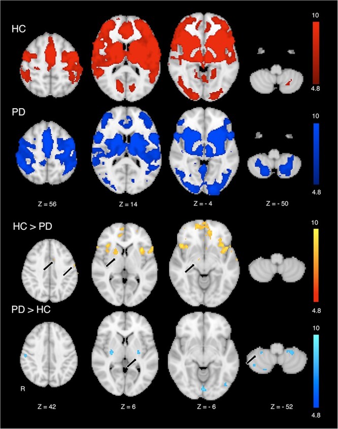
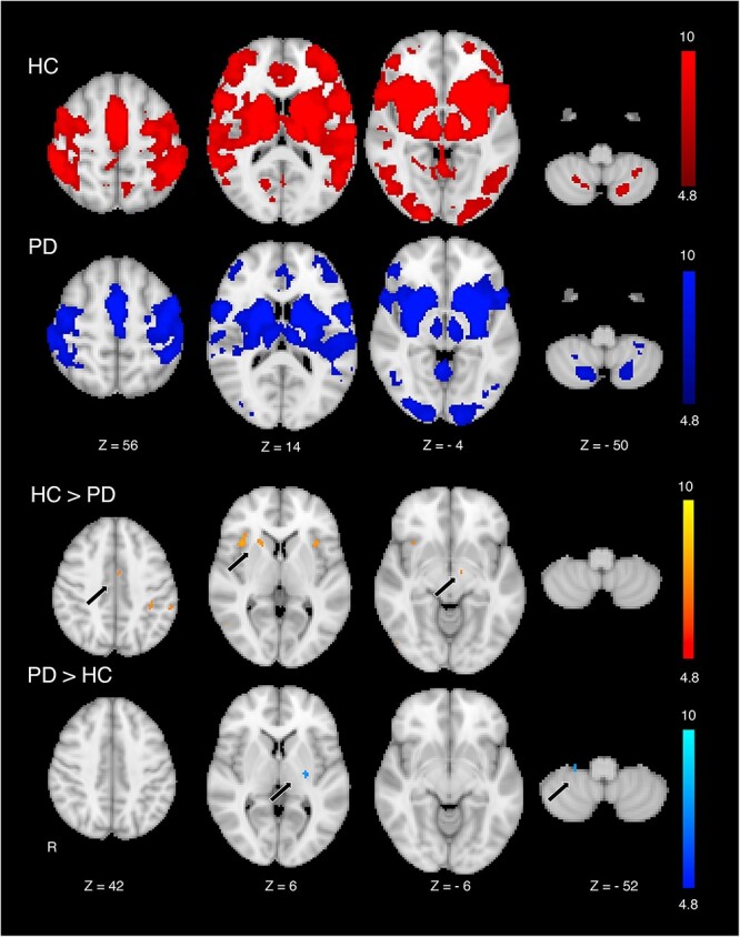
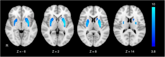
We aimed to clarify whether dopamine depletion in the posterior dorsal striatum in early-stage Parkinson's disease (PD) alters synchronized activity in the cortico-basal ganglia motor circuit. In sum, 14 PD patients and 16 matched healthy controls (HC) underwent [11C]-2-β-carbomethoxy-3-β-(4-fluorophenyl) tropane positron emission tomography to identify striatal dopamine-depleted areas. The identified map was applied to functional magnetic resonance imaging (fMRI) to discover abnormalities in functional connectivity (FC) during motor-task and rest-state in PD patients in the drug-off state relative to HC. Striatal dopamine-depleted areas formed synchronized fMRI activity that largely corresponded to the cortico-basal ganglia motor circuit. Group comparisons revealed that striatal dopamine-depleted areas exhibited decreased FC with the medial premotor cortex during motor-task and with the medial, lateral premotor and primary motor cortices during rest-state. Striatal dopamine-depleted areas also elucidated decreased FC in the subthalamic nucleus (STN) in PD both during motor-task and rest-state. The STN regions that exhibited reduced FC with striatal dopamine-depleted areas demonstrated excessive FC with the lateral premotor and primary motor cortices in PD only during rest-state. Our findings suggest that striatal dopamine-depleted area reduced synchronized activity with the motor cortices and STN, which, in turn, induces an abnormal increase in coupling between the areas in PD.


 分享
分享

 求助内容:
求助内容: 应助结果提醒方式:
 扫码关注我们
