{"title":"GATA3阳性多形性脂肪肉瘤1例,上皮样变异:一个诊断缺陷。","authors":"Makoto Abe, Nobuo Hoshi, Sayuri Hoshi, Kaoru Hirabayashi, Kazutaka Kikuta, Toru Hirozane, Rumi Nakagawa, Tsukasa Mizuno, Hiroshi Nakamura, Koichi Inoue, Takehiko Yamaguchi","doi":"10.1155/2023/9443027","DOIUrl":null,"url":null,"abstract":"<p><p>Pleomorphic liposarcoma is a rare malignant adipocytic tumor showing undifferentiated pleomorphic sarcoma morphology with various degrees of epithelioid features. It is sometimes difficult to distinguish from carcinoma metastasis. Immunohistochemical panel is very important for differential diagnosis; however, there is a risk that unexpected staining could lead to misinterpretation. We report a pleomorphic liposarcoma, epithelioid variant, in an 88-year-old man, with tricky-positive staining for GATA3. Histological examination revealed a tumor with epithelioid morphology. The tumor consists of solid sheets of epithelioid tumor cells with focal aggregates of pleomorphic lipoblasts. Immunohistochemically, the adipocytic tumor cell areas were positive for S100 protein, and the epithelioid tumor cells showed CAM 5.2 positivity. GATA3 was diffusely positive. The combination of CAM 5.2 and GATA3 staining suggested the possibility of metastatic cancer, but systemic clinical examinations did not detect any presence of a primary tumor, including urinary bladder, breasts, and salivary glands. The pathological diagnosis of pleomorphic liposarcoma, epithelioid variant, was made because of the presence of malignant lipoblasts. Our report may contribute for differential diagnosis of pleomorphic liposarcoma, epithelioid variant, with unexpected positive immunoreaction for GATA3.</p>","PeriodicalId":45638,"journal":{"name":"Case Reports in Pathology","volume":"2023 ","pages":"9443027"},"PeriodicalIF":0.7000,"publicationDate":"2023-01-01","publicationTypes":"Journal Article","fieldsOfStudy":null,"isOpenAccess":false,"openAccessPdf":"https://www.ncbi.nlm.nih.gov/pmc/articles/PMC10065854/pdf/","citationCount":"0","resultStr":"{\"title\":\"A Case of GATA3 Positive Pleomorphic Liposarcoma, Epithelioid Variant: A Diagnostic Pitfall.\",\"authors\":\"Makoto Abe, Nobuo Hoshi, Sayuri Hoshi, Kaoru Hirabayashi, Kazutaka Kikuta, Toru Hirozane, Rumi Nakagawa, Tsukasa Mizuno, Hiroshi Nakamura, Koichi Inoue, Takehiko Yamaguchi\",\"doi\":\"10.1155/2023/9443027\",\"DOIUrl\":null,\"url\":null,\"abstract\":\"<p><p>Pleomorphic liposarcoma is a rare malignant adipocytic tumor showing undifferentiated pleomorphic sarcoma morphology with various degrees of epithelioid features. It is sometimes difficult to distinguish from carcinoma metastasis. Immunohistochemical panel is very important for differential diagnosis; however, there is a risk that unexpected staining could lead to misinterpretation. We report a pleomorphic liposarcoma, epithelioid variant, in an 88-year-old man, with tricky-positive staining for GATA3. Histological examination revealed a tumor with epithelioid morphology. The tumor consists of solid sheets of epithelioid tumor cells with focal aggregates of pleomorphic lipoblasts. Immunohistochemically, the adipocytic tumor cell areas were positive for S100 protein, and the epithelioid tumor cells showed CAM 5.2 positivity. GATA3 was diffusely positive. The combination of CAM 5.2 and GATA3 staining suggested the possibility of metastatic cancer, but systemic clinical examinations did not detect any presence of a primary tumor, including urinary bladder, breasts, and salivary glands. The pathological diagnosis of pleomorphic liposarcoma, epithelioid variant, was made because of the presence of malignant lipoblasts. Our report may contribute for differential diagnosis of pleomorphic liposarcoma, epithelioid variant, with unexpected positive immunoreaction for GATA3.</p>\",\"PeriodicalId\":45638,\"journal\":{\"name\":\"Case Reports in Pathology\",\"volume\":\"2023 \",\"pages\":\"9443027\"},\"PeriodicalIF\":0.7000,\"publicationDate\":\"2023-01-01\",\"publicationTypes\":\"Journal Article\",\"fieldsOfStudy\":null,\"isOpenAccess\":false,\"openAccessPdf\":\"https://www.ncbi.nlm.nih.gov/pmc/articles/PMC10065854/pdf/\",\"citationCount\":\"0\",\"resultStr\":null,\"platform\":\"Semanticscholar\",\"paperid\":null,\"PeriodicalName\":\"Case Reports in Pathology\",\"FirstCategoryId\":\"1085\",\"ListUrlMain\":\"https://doi.org/10.1155/2023/9443027\",\"RegionNum\":0,\"RegionCategory\":null,\"ArticlePicture\":[],\"TitleCN\":null,\"AbstractTextCN\":null,\"PMCID\":null,\"EPubDate\":\"\",\"PubModel\":\"\",\"JCR\":\"Q4\",\"JCRName\":\"PATHOLOGY\",\"Score\":null,\"Total\":0}","platform":"Semanticscholar","paperid":null,"PeriodicalName":"Case Reports in Pathology","FirstCategoryId":"1085","ListUrlMain":"https://doi.org/10.1155/2023/9443027","RegionNum":0,"RegionCategory":null,"ArticlePicture":[],"TitleCN":null,"AbstractTextCN":null,"PMCID":null,"EPubDate":"","PubModel":"","JCR":"Q4","JCRName":"PATHOLOGY","Score":null,"Total":0}
A Case of GATA3 Positive Pleomorphic Liposarcoma, Epithelioid Variant: A Diagnostic Pitfall.
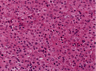
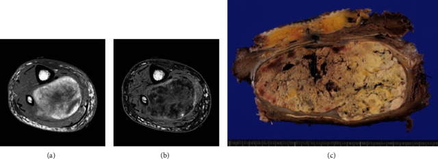
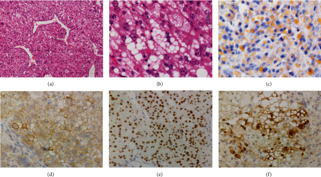
Pleomorphic liposarcoma is a rare malignant adipocytic tumor showing undifferentiated pleomorphic sarcoma morphology with various degrees of epithelioid features. It is sometimes difficult to distinguish from carcinoma metastasis. Immunohistochemical panel is very important for differential diagnosis; however, there is a risk that unexpected staining could lead to misinterpretation. We report a pleomorphic liposarcoma, epithelioid variant, in an 88-year-old man, with tricky-positive staining for GATA3. Histological examination revealed a tumor with epithelioid morphology. The tumor consists of solid sheets of epithelioid tumor cells with focal aggregates of pleomorphic lipoblasts. Immunohistochemically, the adipocytic tumor cell areas were positive for S100 protein, and the epithelioid tumor cells showed CAM 5.2 positivity. GATA3 was diffusely positive. The combination of CAM 5.2 and GATA3 staining suggested the possibility of metastatic cancer, but systemic clinical examinations did not detect any presence of a primary tumor, including urinary bladder, breasts, and salivary glands. The pathological diagnosis of pleomorphic liposarcoma, epithelioid variant, was made because of the presence of malignant lipoblasts. Our report may contribute for differential diagnosis of pleomorphic liposarcoma, epithelioid variant, with unexpected positive immunoreaction for GATA3.


 分享
分享
 求助内容:
求助内容: 应助结果提醒方式:
 扫码关注我们
