{"title":"COP1通过PI3K/AKT信号通路下调CDH18促进胃癌发生的作用","authors":"Benhuo Zhao, Jiaojiao Wu, Xiuli Cha, Guangtong Mao, Hengliang Shi, Sujuan Fei, Bei Miao","doi":"10.1155/2023/5617875","DOIUrl":null,"url":null,"abstract":"<p><p>In recent years, the involvement of E3 ubiquitin ligase constitutive photomorphogenesis 1 (COP1) in the tumorigenesis of gastric cancer (GC) has been elucidated. However, the exact underlying mechanism remains to be clarified. In the present study, the expression profiles of COP1 in GC were derived from the Gene Expression Omnibus (GEO) and the Cancer Genome Atlas (TCGA) databases, followed by verification via immunohistochemical staining (IHC), Western blotting (WB), and quantitative real-time polymerase chain reaction (qRT-PCR) reaction assays on clinical samples. In vitro, the gain- and loss-of-function experiments of COP1 protein were conducted to explore its role in GC cell lines HGC-27 and SGC-7901. Furthermore, we screened the interaction protein of COP1 by yeast two-hybrid experiment and verified their combination by co-immunoprecipitation (co-IP). We preliminary explored the possible underlying mechanisms of COP1 protein in GC cell lines via WB. COP1 was upregulated in GC tissues compared with the corresponding non-carcinoma tissues. In vitro, the upregulation of COP1 protein promoted the proliferation and migration of GC cells. The yeast two-hybrid experiment and co-IP indicated that Cadherin 18 (CDH18) could constitute a complex with COP1. Moreover, cells with COP1 over-expression showed low levels of CDH18 expression, with the intracellular PI3K/AKT pathway activated and the malignancy of GC cell lines enhanced. Our findings demonstrated that COP1 promoted the GC tumorigenesis by downregulated CDH18 with the involvement of PI3K/AKT signaling pathway in cell lines, suggesting the potential of COP1 as a prognostic biomarker and therapeutic target for GC.</p>","PeriodicalId":49326,"journal":{"name":"Analytical Cellular Pathology","volume":"2023 ","pages":"5617875"},"PeriodicalIF":2.7000,"publicationDate":"2023-01-01","publicationTypes":"Journal Article","fieldsOfStudy":null,"isOpenAccess":false,"openAccessPdf":"https://www.ncbi.nlm.nih.gov/pmc/articles/PMC10072965/pdf/","citationCount":"0","resultStr":"{\"title\":\"Effect of COP1 in Promoting the Tumorigenesis of Gastric Cancer by Down-Regulation of CDH18 via PI3K/AKT Signal Pathway.\",\"authors\":\"Benhuo Zhao, Jiaojiao Wu, Xiuli Cha, Guangtong Mao, Hengliang Shi, Sujuan Fei, Bei Miao\",\"doi\":\"10.1155/2023/5617875\",\"DOIUrl\":null,\"url\":null,\"abstract\":\"<p><p>In recent years, the involvement of E3 ubiquitin ligase constitutive photomorphogenesis 1 (COP1) in the tumorigenesis of gastric cancer (GC) has been elucidated. However, the exact underlying mechanism remains to be clarified. In the present study, the expression profiles of COP1 in GC were derived from the Gene Expression Omnibus (GEO) and the Cancer Genome Atlas (TCGA) databases, followed by verification via immunohistochemical staining (IHC), Western blotting (WB), and quantitative real-time polymerase chain reaction (qRT-PCR) reaction assays on clinical samples. In vitro, the gain- and loss-of-function experiments of COP1 protein were conducted to explore its role in GC cell lines HGC-27 and SGC-7901. Furthermore, we screened the interaction protein of COP1 by yeast two-hybrid experiment and verified their combination by co-immunoprecipitation (co-IP). We preliminary explored the possible underlying mechanisms of COP1 protein in GC cell lines via WB. COP1 was upregulated in GC tissues compared with the corresponding non-carcinoma tissues. In vitro, the upregulation of COP1 protein promoted the proliferation and migration of GC cells. The yeast two-hybrid experiment and co-IP indicated that Cadherin 18 (CDH18) could constitute a complex with COP1. Moreover, cells with COP1 over-expression showed low levels of CDH18 expression, with the intracellular PI3K/AKT pathway activated and the malignancy of GC cell lines enhanced. Our findings demonstrated that COP1 promoted the GC tumorigenesis by downregulated CDH18 with the involvement of PI3K/AKT signaling pathway in cell lines, suggesting the potential of COP1 as a prognostic biomarker and therapeutic target for GC.</p>\",\"PeriodicalId\":49326,\"journal\":{\"name\":\"Analytical Cellular Pathology\",\"volume\":\"2023 \",\"pages\":\"5617875\"},\"PeriodicalIF\":2.7000,\"publicationDate\":\"2023-01-01\",\"publicationTypes\":\"Journal Article\",\"fieldsOfStudy\":null,\"isOpenAccess\":false,\"openAccessPdf\":\"https://www.ncbi.nlm.nih.gov/pmc/articles/PMC10072965/pdf/\",\"citationCount\":\"0\",\"resultStr\":null,\"platform\":\"Semanticscholar\",\"paperid\":null,\"PeriodicalName\":\"Analytical Cellular Pathology\",\"FirstCategoryId\":\"3\",\"ListUrlMain\":\"https://doi.org/10.1155/2023/5617875\",\"RegionNum\":4,\"RegionCategory\":\"医学\",\"ArticlePicture\":[],\"TitleCN\":null,\"AbstractTextCN\":null,\"PMCID\":null,\"EPubDate\":\"\",\"PubModel\":\"\",\"JCR\":\"Q3\",\"JCRName\":\"CELL BIOLOGY\",\"Score\":null,\"Total\":0}","platform":"Semanticscholar","paperid":null,"PeriodicalName":"Analytical Cellular Pathology","FirstCategoryId":"3","ListUrlMain":"https://doi.org/10.1155/2023/5617875","RegionNum":4,"RegionCategory":"医学","ArticlePicture":[],"TitleCN":null,"AbstractTextCN":null,"PMCID":null,"EPubDate":"","PubModel":"","JCR":"Q3","JCRName":"CELL BIOLOGY","Score":null,"Total":0}
Effect of COP1 in Promoting the Tumorigenesis of Gastric Cancer by Down-Regulation of CDH18 via PI3K/AKT Signal Pathway.
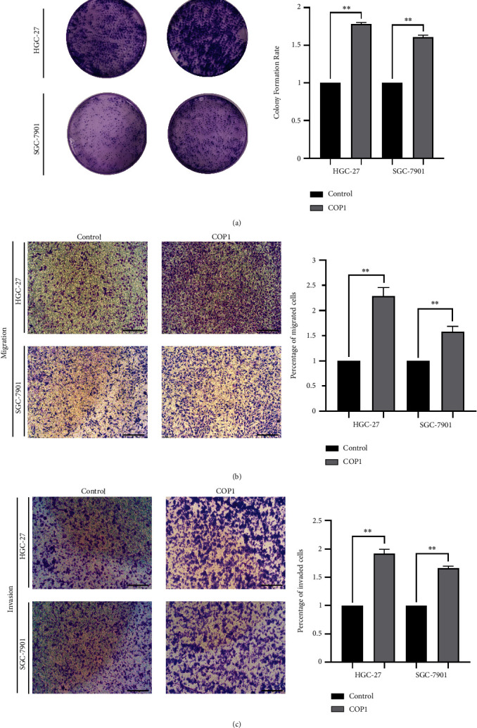
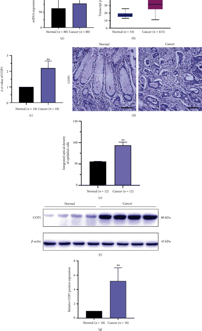
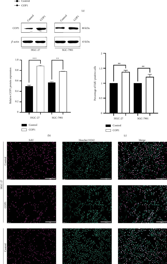
In recent years, the involvement of E3 ubiquitin ligase constitutive photomorphogenesis 1 (COP1) in the tumorigenesis of gastric cancer (GC) has been elucidated. However, the exact underlying mechanism remains to be clarified. In the present study, the expression profiles of COP1 in GC were derived from the Gene Expression Omnibus (GEO) and the Cancer Genome Atlas (TCGA) databases, followed by verification via immunohistochemical staining (IHC), Western blotting (WB), and quantitative real-time polymerase chain reaction (qRT-PCR) reaction assays on clinical samples. In vitro, the gain- and loss-of-function experiments of COP1 protein were conducted to explore its role in GC cell lines HGC-27 and SGC-7901. Furthermore, we screened the interaction protein of COP1 by yeast two-hybrid experiment and verified their combination by co-immunoprecipitation (co-IP). We preliminary explored the possible underlying mechanisms of COP1 protein in GC cell lines via WB. COP1 was upregulated in GC tissues compared with the corresponding non-carcinoma tissues. In vitro, the upregulation of COP1 protein promoted the proliferation and migration of GC cells. The yeast two-hybrid experiment and co-IP indicated that Cadherin 18 (CDH18) could constitute a complex with COP1. Moreover, cells with COP1 over-expression showed low levels of CDH18 expression, with the intracellular PI3K/AKT pathway activated and the malignancy of GC cell lines enhanced. Our findings demonstrated that COP1 promoted the GC tumorigenesis by downregulated CDH18 with the involvement of PI3K/AKT signaling pathway in cell lines, suggesting the potential of COP1 as a prognostic biomarker and therapeutic target for GC.
期刊介绍:
Analytical Cellular Pathology is a peer-reviewed, Open Access journal that provides a forum for scientists, medical practitioners and pathologists working in the area of cellular pathology. The journal publishes original research articles, review articles, and clinical studies related to cytology, carcinogenesis, cell receptors, biomarkers, diagnostic pathology, immunopathology, and hematology.


 分享
分享
 求助内容:
求助内容: 应助结果提醒方式:
 扫码关注我们
