{"title":"线性探头超声诊断眶下缘骨折的准确性。","authors":"Chatchai Pruksapong, Nuttadon Wongprakob, Minth Panphichet","doi":"10.1186/s13089-022-00298-y","DOIUrl":null,"url":null,"abstract":"<p><strong>Background: </strong>Maxillofacial fractures are a common cause of visits to emergency department, accounting for more than 400,000 annual visits in the United States. Gold standard diagnostic tool is conventional computerized tomography (CT) or 3DCT reconstruction. However, the disadvantages of CT are radiation exposure, unavailable in some hospital and expensiveness. Whereas the bony structures overlap is a problem in diagnostic when using plain film X-ray. The objective of this study is to show the accuracy of a linear-probe ultrasound compared to computed tomography and plain film X-ray in diagnosis of infraorbital rim fracture.</p><p><strong>Methods: </strong>Patients clinically suspected of an inferior orbital rim fracture underwent linear-probe ultrasonographic investigation, plain film X-ray and CT. CT was used as gold standard in this diagnostic study. A radiologist and senior resident of plastic surgery were the examiner and interobserver for comparison.</p><p><strong>Result: </strong>A total of 34 patients with suspected infraorbital rim fractures were investigated. Sensitivity of the linear-probe ultrasonography versus CT in the detection of infraorbital rim fracture was 92.9% (95% CI 66.1-99.8), specificity was 90.0% (95% CI 68.3-98.8), positive predictive value was 86.7% (95% CI 59.5-98.3), negative predictive value was 94.7% ( 95% CI 74.0-99.9), accuracy 91%.</p><p><strong>Conclusion: </strong>Linear probe ultrasonography is a good diagnostic tool and has better reliability than the plain film X-ray and can be used as alternative to CT in inferior orbital rim fracture.</p>","PeriodicalId":75201,"journal":{"name":"","volume":"15 1","pages":"9"},"PeriodicalIF":0.0,"publicationDate":"2023-02-10","publicationTypes":"Journal Article","fieldsOfStudy":null,"isOpenAccess":false,"openAccessPdf":"https://www.ncbi.nlm.nih.gov/pmc/articles/PMC9918656/pdf/","citationCount":"0","resultStr":"{\"title\":\"Accuracy of linear-probe ultrasonography in diagnosis of infraorbital rim fractures.\",\"authors\":\"Chatchai Pruksapong, Nuttadon Wongprakob, Minth Panphichet\",\"doi\":\"10.1186/s13089-022-00298-y\",\"DOIUrl\":null,\"url\":null,\"abstract\":\"<p><strong>Background: </strong>Maxillofacial fractures are a common cause of visits to emergency department, accounting for more than 400,000 annual visits in the United States. Gold standard diagnostic tool is conventional computerized tomography (CT) or 3DCT reconstruction. However, the disadvantages of CT are radiation exposure, unavailable in some hospital and expensiveness. Whereas the bony structures overlap is a problem in diagnostic when using plain film X-ray. The objective of this study is to show the accuracy of a linear-probe ultrasound compared to computed tomography and plain film X-ray in diagnosis of infraorbital rim fracture.</p><p><strong>Methods: </strong>Patients clinically suspected of an inferior orbital rim fracture underwent linear-probe ultrasonographic investigation, plain film X-ray and CT. CT was used as gold standard in this diagnostic study. A radiologist and senior resident of plastic surgery were the examiner and interobserver for comparison.</p><p><strong>Result: </strong>A total of 34 patients with suspected infraorbital rim fractures were investigated. Sensitivity of the linear-probe ultrasonography versus CT in the detection of infraorbital rim fracture was 92.9% (95% CI 66.1-99.8), specificity was 90.0% (95% CI 68.3-98.8), positive predictive value was 86.7% (95% CI 59.5-98.3), negative predictive value was 94.7% ( 95% CI 74.0-99.9), accuracy 91%.</p><p><strong>Conclusion: </strong>Linear probe ultrasonography is a good diagnostic tool and has better reliability than the plain film X-ray and can be used as alternative to CT in inferior orbital rim fracture.</p>\",\"PeriodicalId\":75201,\"journal\":{\"name\":\"\",\"volume\":\"15 1\",\"pages\":\"9\"},\"PeriodicalIF\":0.0,\"publicationDate\":\"2023-02-10\",\"publicationTypes\":\"Journal Article\",\"fieldsOfStudy\":null,\"isOpenAccess\":false,\"openAccessPdf\":\"https://www.ncbi.nlm.nih.gov/pmc/articles/PMC9918656/pdf/\",\"citationCount\":\"0\",\"resultStr\":null,\"platform\":\"Semanticscholar\",\"paperid\":null,\"PeriodicalName\":\"\",\"FirstCategoryId\":\"1085\",\"ListUrlMain\":\"https://doi.org/10.1186/s13089-022-00298-y\",\"RegionNum\":0,\"RegionCategory\":null,\"ArticlePicture\":[],\"TitleCN\":null,\"AbstractTextCN\":null,\"PMCID\":null,\"EPubDate\":\"\",\"PubModel\":\"\",\"JCR\":\"\",\"JCRName\":\"\",\"Score\":null,\"Total\":0}","platform":"Semanticscholar","paperid":null,"PeriodicalName":"","FirstCategoryId":"1085","ListUrlMain":"https://doi.org/10.1186/s13089-022-00298-y","RegionNum":0,"RegionCategory":null,"ArticlePicture":[],"TitleCN":null,"AbstractTextCN":null,"PMCID":null,"EPubDate":"","PubModel":"","JCR":"","JCRName":"","Score":null,"Total":0}
引用次数: 0
摘要
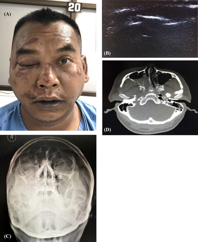
背景:颌面部骨折是急诊科就诊的常见原因,在美国每年有超过40万人次就诊。金标准诊断工具是传统的计算机断层扫描(CT)或3DCT重建。然而,CT的缺点是有辐射,一些医院没有,而且价格昂贵。而x线平片诊断时,骨结构重叠是一个问题。本研究的目的是显示线性探针超声诊断眶下缘骨折的准确性,与计算机断层扫描和x光平片相比。方法:对临床怀疑眶下缘骨折的患者行线性探头超声、x线平片及CT检查。CT作为本诊断研究的金标准。一名放射科医师和一名整形外科资深住院医师作为审查员和相互观察者进行比较。结果:对34例疑似眶下缘骨折患者进行了调查。线性探头超声与CT检测眶下缘骨折的敏感性为92.9% (95% CI 66.1-99.8),特异性为90.0% (95% CI 68.3-98.8),阳性预测值为86.7% (95% CI 59.5-98.3),阴性预测值为94.7% (95% CI 74.0-99.9),准确率为91%。结论:线性探头超声是一种较好的诊断工具,可靠性优于x线平片,可替代CT诊断下眶缘骨折。
Accuracy of linear-probe ultrasonography in diagnosis of infraorbital rim fractures.
Background: Maxillofacial fractures are a common cause of visits to emergency department, accounting for more than 400,000 annual visits in the United States. Gold standard diagnostic tool is conventional computerized tomography (CT) or 3DCT reconstruction. However, the disadvantages of CT are radiation exposure, unavailable in some hospital and expensiveness. Whereas the bony structures overlap is a problem in diagnostic when using plain film X-ray. The objective of this study is to show the accuracy of a linear-probe ultrasound compared to computed tomography and plain film X-ray in diagnosis of infraorbital rim fracture.
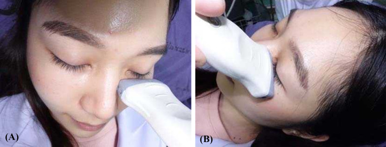
Methods: Patients clinically suspected of an inferior orbital rim fracture underwent linear-probe ultrasonographic investigation, plain film X-ray and CT. CT was used as gold standard in this diagnostic study. A radiologist and senior resident of plastic surgery were the examiner and interobserver for comparison.
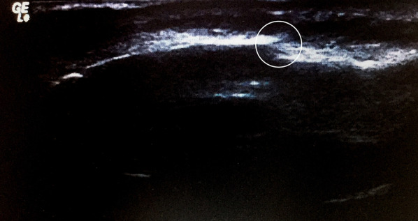
Result: A total of 34 patients with suspected infraorbital rim fractures were investigated. Sensitivity of the linear-probe ultrasonography versus CT in the detection of infraorbital rim fracture was 92.9% (95% CI 66.1-99.8), specificity was 90.0% (95% CI 68.3-98.8), positive predictive value was 86.7% (95% CI 59.5-98.3), negative predictive value was 94.7% ( 95% CI 74.0-99.9), accuracy 91%.
Conclusion: Linear probe ultrasonography is a good diagnostic tool and has better reliability than the plain film X-ray and can be used as alternative to CT in inferior orbital rim fracture.


 分享
分享
 求助内容:
求助内容: 应助结果提醒方式:
 扫码关注我们
