{"title":"脑脊液对预防脑血栓栓塞和血脑屏障破坏的意外作用:首次实验研究。","authors":"Mete Zeynal","doi":"10.5152/eurasianjmed.2023.22317","DOIUrl":null,"url":null,"abstract":"<p><strong>Objective: </strong>We investigated the presence of thromboembolism that may develop in hippocampal arteries due to decreased cerebrospinal fluid volume because of choroid plexus damage caused by subarachnoid hemorrhage.</p><p><strong>Materials and methods: </strong>Twenty-four rabbits were included as test subjects in this study. The study group comprised 14 test subjects administered autologous blood (0.5 mL). Coronary sections of the temporal uncus were prepared to observe the choroid plexus and the hippocampus together. Cellular shrinkage, darkening, halo formation, and ciliary element loss were considered criteria for degeneration. Blood-brain barriers were also examined in the hippocampus. The density of degenerated epithelial cells in the choroid plexus (n/mm3 ) and thromboembolisms in the hippocampal arteries (n/cm2 ) were compared statistically.</p><p><strong>Results: </strong>Histopathological examination revealed that the number of degenerated epithelial cells in the choroid plexus and the number of thromboembolisms in the hippocampal arteries were 7 ± 2 and 1 ± 1 in group 1, 16 ± 4 and 3 ± 1 in group 2, and 64 ± 9 and 6 ± 2 in group 3, respectively. The significance levels were P < .005 for group 1 vs. group 2, P < .0005 for group 2 vs. group 3, and P < .00001 for group 1 vs. group 3.</p><p><strong>Conclusion: </strong>This study demonstrates that decreased cerebrospinal fluid volume induced by choroid plexus degeneration causes cerebral thromboembolism following subarachnoid hemorrhage, which has not been previously described.</p>","PeriodicalId":74999,"journal":{"name":"","volume":"55 1","pages":"50-53"},"PeriodicalIF":0.0,"publicationDate":"2023-02-01","publicationTypes":"Journal Article","fieldsOfStudy":null,"isOpenAccess":false,"openAccessPdf":"https://www.ncbi.nlm.nih.gov/pmc/articles/PMC10081128/pdf/","citationCount":"0","resultStr":"{\"title\":\"Unexpected Effects of Cerebrospinal Fluid on the Prevention of Cerebral Thromboembolism and Blood-Brain Barrier Disruption: First Experimental Study.\",\"authors\":\"Mete Zeynal\",\"doi\":\"10.5152/eurasianjmed.2023.22317\",\"DOIUrl\":null,\"url\":null,\"abstract\":\"<p><strong>Objective: </strong>We investigated the presence of thromboembolism that may develop in hippocampal arteries due to decreased cerebrospinal fluid volume because of choroid plexus damage caused by subarachnoid hemorrhage.</p><p><strong>Materials and methods: </strong>Twenty-four rabbits were included as test subjects in this study. The study group comprised 14 test subjects administered autologous blood (0.5 mL). Coronary sections of the temporal uncus were prepared to observe the choroid plexus and the hippocampus together. Cellular shrinkage, darkening, halo formation, and ciliary element loss were considered criteria for degeneration. Blood-brain barriers were also examined in the hippocampus. The density of degenerated epithelial cells in the choroid plexus (n/mm3 ) and thromboembolisms in the hippocampal arteries (n/cm2 ) were compared statistically.</p><p><strong>Results: </strong>Histopathological examination revealed that the number of degenerated epithelial cells in the choroid plexus and the number of thromboembolisms in the hippocampal arteries were 7 ± 2 and 1 ± 1 in group 1, 16 ± 4 and 3 ± 1 in group 2, and 64 ± 9 and 6 ± 2 in group 3, respectively. The significance levels were P < .005 for group 1 vs. group 2, P < .0005 for group 2 vs. group 3, and P < .00001 for group 1 vs. group 3.</p><p><strong>Conclusion: </strong>This study demonstrates that decreased cerebrospinal fluid volume induced by choroid plexus degeneration causes cerebral thromboembolism following subarachnoid hemorrhage, which has not been previously described.</p>\",\"PeriodicalId\":74999,\"journal\":{\"name\":\"\",\"volume\":\"55 1\",\"pages\":\"50-53\"},\"PeriodicalIF\":0.0,\"publicationDate\":\"2023-02-01\",\"publicationTypes\":\"Journal Article\",\"fieldsOfStudy\":null,\"isOpenAccess\":false,\"openAccessPdf\":\"https://www.ncbi.nlm.nih.gov/pmc/articles/PMC10081128/pdf/\",\"citationCount\":\"0\",\"resultStr\":null,\"platform\":\"Semanticscholar\",\"paperid\":null,\"PeriodicalName\":\"\",\"FirstCategoryId\":\"1085\",\"ListUrlMain\":\"https://doi.org/10.5152/eurasianjmed.2023.22317\",\"RegionNum\":0,\"RegionCategory\":null,\"ArticlePicture\":[],\"TitleCN\":null,\"AbstractTextCN\":null,\"PMCID\":null,\"EPubDate\":\"\",\"PubModel\":\"\",\"JCR\":\"\",\"JCRName\":\"\",\"Score\":null,\"Total\":0}","platform":"Semanticscholar","paperid":null,"PeriodicalName":"","FirstCategoryId":"1085","ListUrlMain":"https://doi.org/10.5152/eurasianjmed.2023.22317","RegionNum":0,"RegionCategory":null,"ArticlePicture":[],"TitleCN":null,"AbstractTextCN":null,"PMCID":null,"EPubDate":"","PubModel":"","JCR":"","JCRName":"","Score":null,"Total":0}
Unexpected Effects of Cerebrospinal Fluid on the Prevention of Cerebral Thromboembolism and Blood-Brain Barrier Disruption: First Experimental Study.
Objective: We investigated the presence of thromboembolism that may develop in hippocampal arteries due to decreased cerebrospinal fluid volume because of choroid plexus damage caused by subarachnoid hemorrhage.
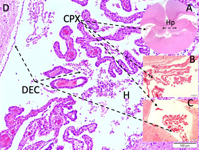
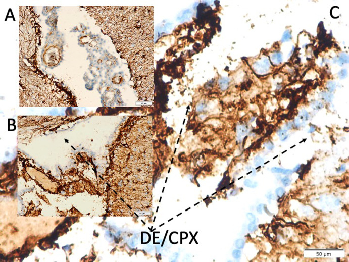
Materials and methods: Twenty-four rabbits were included as test subjects in this study. The study group comprised 14 test subjects administered autologous blood (0.5 mL). Coronary sections of the temporal uncus were prepared to observe the choroid plexus and the hippocampus together. Cellular shrinkage, darkening, halo formation, and ciliary element loss were considered criteria for degeneration. Blood-brain barriers were also examined in the hippocampus. The density of degenerated epithelial cells in the choroid plexus (n/mm3 ) and thromboembolisms in the hippocampal arteries (n/cm2 ) were compared statistically.
Results: Histopathological examination revealed that the number of degenerated epithelial cells in the choroid plexus and the number of thromboembolisms in the hippocampal arteries were 7 ± 2 and 1 ± 1 in group 1, 16 ± 4 and 3 ± 1 in group 2, and 64 ± 9 and 6 ± 2 in group 3, respectively. The significance levels were P < .005 for group 1 vs. group 2, P < .0005 for group 2 vs. group 3, and P < .00001 for group 1 vs. group 3.
Conclusion: This study demonstrates that decreased cerebrospinal fluid volume induced by choroid plexus degeneration causes cerebral thromboembolism following subarachnoid hemorrhage, which has not been previously described.


 分享
分享
 求助内容:
求助内容: 应助结果提醒方式:
 扫码关注我们
