Sajjad Saghebdoust, Mohammad Amin Habibi, Mehran Ekrami, Farshid Khadivar, Mohammad Moein Vakilzadeh, Reza Zare
{"title":"颞叶实质内神经鞘瘤1例报告及文献复习。","authors":"Sajjad Saghebdoust, Mohammad Amin Habibi, Mehran Ekrami, Farshid Khadivar, Mohammad Moein Vakilzadeh, Reza Zare","doi":"10.1055/s-0043-1763525","DOIUrl":null,"url":null,"abstract":"<p><p>Intracranial schwannomas (ISs) account for approximately 8% of intracranial tumors, while IS, a rare entity, is responsible for roughly 1% of IS. A 33-year-old man with a 3-month headache and sudden onset seizure was referred to our clinic. Preoperative magnetic resonance imaging revealed a contrast-enhancing mass accompanied by cystic components in the right temporal lobe. Ganglioglioma, metastasis, or glioblastoma multiforme was suspected, and surgery was advised. During surgery, gross total resection of a noninvasive tumor was conducted. Postoperative recovery was uneventful. Based on histopathological examination and confirmatory immunohistochemistry, the intraparenchymal temporal tumor was diagnosed as schwannoma. ISs are extremely scarce brain tumors mainly located on the surface of the brain or adjacent brain ventricles. The definite preoperative diagnosis of schwannoma cannot be readily established due to radiologically indistinguishable features from metastasis and gliomas; however, histopathology and immunohistochemistry are of great assistance. Complete surgical removal is the most preferred treatment alternative with a long-term favorable prognosis without adjuvant and neoadjuvant chemotherapy requirements.</p>","PeriodicalId":8521,"journal":{"name":"Asian Journal of Neurosurgery","volume":"18 1","pages":"191-195"},"PeriodicalIF":0.0000,"publicationDate":"2023-03-01","publicationTypes":"Journal Article","fieldsOfStudy":null,"isOpenAccess":false,"openAccessPdf":"https://ftp.ncbi.nlm.nih.gov/pub/pmc/oa_pdf/c4/1a/10-1055-s-0043-1763525.PMC10089735.pdf","citationCount":"0","resultStr":"{\"title\":\"Intraparenchymal Schwannoma of Temporal Lobe: A Case Report and Review of the Literature.\",\"authors\":\"Sajjad Saghebdoust, Mohammad Amin Habibi, Mehran Ekrami, Farshid Khadivar, Mohammad Moein Vakilzadeh, Reza Zare\",\"doi\":\"10.1055/s-0043-1763525\",\"DOIUrl\":null,\"url\":null,\"abstract\":\"<p><p>Intracranial schwannomas (ISs) account for approximately 8% of intracranial tumors, while IS, a rare entity, is responsible for roughly 1% of IS. A 33-year-old man with a 3-month headache and sudden onset seizure was referred to our clinic. Preoperative magnetic resonance imaging revealed a contrast-enhancing mass accompanied by cystic components in the right temporal lobe. Ganglioglioma, metastasis, or glioblastoma multiforme was suspected, and surgery was advised. During surgery, gross total resection of a noninvasive tumor was conducted. Postoperative recovery was uneventful. Based on histopathological examination and confirmatory immunohistochemistry, the intraparenchymal temporal tumor was diagnosed as schwannoma. ISs are extremely scarce brain tumors mainly located on the surface of the brain or adjacent brain ventricles. The definite preoperative diagnosis of schwannoma cannot be readily established due to radiologically indistinguishable features from metastasis and gliomas; however, histopathology and immunohistochemistry are of great assistance. Complete surgical removal is the most preferred treatment alternative with a long-term favorable prognosis without adjuvant and neoadjuvant chemotherapy requirements.</p>\",\"PeriodicalId\":8521,\"journal\":{\"name\":\"Asian Journal of Neurosurgery\",\"volume\":\"18 1\",\"pages\":\"191-195\"},\"PeriodicalIF\":0.0000,\"publicationDate\":\"2023-03-01\",\"publicationTypes\":\"Journal Article\",\"fieldsOfStudy\":null,\"isOpenAccess\":false,\"openAccessPdf\":\"https://ftp.ncbi.nlm.nih.gov/pub/pmc/oa_pdf/c4/1a/10-1055-s-0043-1763525.PMC10089735.pdf\",\"citationCount\":\"0\",\"resultStr\":null,\"platform\":\"Semanticscholar\",\"paperid\":null,\"PeriodicalName\":\"Asian Journal of Neurosurgery\",\"FirstCategoryId\":\"1085\",\"ListUrlMain\":\"https://doi.org/10.1055/s-0043-1763525\",\"RegionNum\":0,\"RegionCategory\":null,\"ArticlePicture\":[],\"TitleCN\":null,\"AbstractTextCN\":null,\"PMCID\":null,\"EPubDate\":\"\",\"PubModel\":\"\",\"JCR\":\"\",\"JCRName\":\"\",\"Score\":null,\"Total\":0}","platform":"Semanticscholar","paperid":null,"PeriodicalName":"Asian Journal of Neurosurgery","FirstCategoryId":"1085","ListUrlMain":"https://doi.org/10.1055/s-0043-1763525","RegionNum":0,"RegionCategory":null,"ArticlePicture":[],"TitleCN":null,"AbstractTextCN":null,"PMCID":null,"EPubDate":"","PubModel":"","JCR":"","JCRName":"","Score":null,"Total":0}
Intraparenchymal Schwannoma of Temporal Lobe: A Case Report and Review of the Literature.
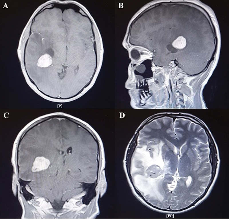
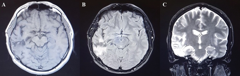
Intracranial schwannomas (ISs) account for approximately 8% of intracranial tumors, while IS, a rare entity, is responsible for roughly 1% of IS. A 33-year-old man with a 3-month headache and sudden onset seizure was referred to our clinic. Preoperative magnetic resonance imaging revealed a contrast-enhancing mass accompanied by cystic components in the right temporal lobe. Ganglioglioma, metastasis, or glioblastoma multiforme was suspected, and surgery was advised. During surgery, gross total resection of a noninvasive tumor was conducted. Postoperative recovery was uneventful. Based on histopathological examination and confirmatory immunohistochemistry, the intraparenchymal temporal tumor was diagnosed as schwannoma. ISs are extremely scarce brain tumors mainly located on the surface of the brain or adjacent brain ventricles. The definite preoperative diagnosis of schwannoma cannot be readily established due to radiologically indistinguishable features from metastasis and gliomas; however, histopathology and immunohistochemistry are of great assistance. Complete surgical removal is the most preferred treatment alternative with a long-term favorable prognosis without adjuvant and neoadjuvant chemotherapy requirements.


 分享
分享

 求助内容:
求助内容: 应助结果提醒方式:
 扫码关注我们
