Giosué DiPellegrini, Riccardo Boccaletti, Anna Mingozzi, Antonella Fara, Domenico Policicchio
{"title":"模拟脑肿瘤的脑弓形虫病的单一丘脑定位:放射学和临床表现。","authors":"Giosué DiPellegrini, Riccardo Boccaletti, Anna Mingozzi, Antonella Fara, Domenico Policicchio","doi":"10.25259/SNI_34_2023","DOIUrl":null,"url":null,"abstract":"Background: Cerebral toxoplasmosis is a relatively rare disorder that usually affects immunocompromised patients. The most common scenario occurs among human immunodeficiency virus (HIV)-positive patients. In those patients, toxoplasmosis is the most frequent cause of expansive brain lesion and continues to cause elevated morbidity and mortality. In typical cases of toxoplasmosis, both computed tomography and magnetic resonance imaging reveal single/ multiple nodular or ring-enhancing lesions with surrounding edema. Nevertheless, cases of cerebral toxoplasmosis with atypical radiological features have been reported. Diagnosis can be obtained by finding organisms in the cerebrospinal fluid or in stereotactic biopsy samples of the brain lesion. If untreated, cerebral toxoplasmosis is uniformly fatal, so prompt diagnosis is mandatory. A prompt diagnosis is necessary, as untreated cerebral toxoplasmosis is uniformly fatal. Case Description: We discuss imaging and clinical findings of a patient – not aware of being HIV-positive – with a solitary atypical brain localization of toxoplasmosis mimicking a brain tumor. Conclusion: Although relatively uncommon, neurosurgeons should be aware of the potential occurrence of cerebral toxoplasmosis. High index of suspicion is needed for timely diagnosis and prompt initiation of therapy.","PeriodicalId":38981,"journal":{"name":"Surgical Neurology International","volume":"14 ","pages":"82"},"PeriodicalIF":0.0000,"publicationDate":"2023-01-01","publicationTypes":"Journal Article","fieldsOfStudy":null,"isOpenAccess":false,"openAccessPdf":"https://ftp.ncbi.nlm.nih.gov/pub/pmc/oa_pdf/c0/33/SNI-14-82.PMC10070335.pdf","citationCount":"0","resultStr":"{\"title\":\"Single thalamic localization of brain toxoplasmosis mimicking brain tumors: Radiological and clinical findings.\",\"authors\":\"Giosué DiPellegrini, Riccardo Boccaletti, Anna Mingozzi, Antonella Fara, Domenico Policicchio\",\"doi\":\"10.25259/SNI_34_2023\",\"DOIUrl\":null,\"url\":null,\"abstract\":\"Background: Cerebral toxoplasmosis is a relatively rare disorder that usually affects immunocompromised patients. The most common scenario occurs among human immunodeficiency virus (HIV)-positive patients. In those patients, toxoplasmosis is the most frequent cause of expansive brain lesion and continues to cause elevated morbidity and mortality. In typical cases of toxoplasmosis, both computed tomography and magnetic resonance imaging reveal single/ multiple nodular or ring-enhancing lesions with surrounding edema. Nevertheless, cases of cerebral toxoplasmosis with atypical radiological features have been reported. Diagnosis can be obtained by finding organisms in the cerebrospinal fluid or in stereotactic biopsy samples of the brain lesion. If untreated, cerebral toxoplasmosis is uniformly fatal, so prompt diagnosis is mandatory. A prompt diagnosis is necessary, as untreated cerebral toxoplasmosis is uniformly fatal. Case Description: We discuss imaging and clinical findings of a patient – not aware of being HIV-positive – with a solitary atypical brain localization of toxoplasmosis mimicking a brain tumor. Conclusion: Although relatively uncommon, neurosurgeons should be aware of the potential occurrence of cerebral toxoplasmosis. High index of suspicion is needed for timely diagnosis and prompt initiation of therapy.\",\"PeriodicalId\":38981,\"journal\":{\"name\":\"Surgical Neurology International\",\"volume\":\"14 \",\"pages\":\"82\"},\"PeriodicalIF\":0.0000,\"publicationDate\":\"2023-01-01\",\"publicationTypes\":\"Journal Article\",\"fieldsOfStudy\":null,\"isOpenAccess\":false,\"openAccessPdf\":\"https://ftp.ncbi.nlm.nih.gov/pub/pmc/oa_pdf/c0/33/SNI-14-82.PMC10070335.pdf\",\"citationCount\":\"0\",\"resultStr\":null,\"platform\":\"Semanticscholar\",\"paperid\":null,\"PeriodicalName\":\"Surgical Neurology International\",\"FirstCategoryId\":\"1085\",\"ListUrlMain\":\"https://doi.org/10.25259/SNI_34_2023\",\"RegionNum\":0,\"RegionCategory\":null,\"ArticlePicture\":[],\"TitleCN\":null,\"AbstractTextCN\":null,\"PMCID\":null,\"EPubDate\":\"\",\"PubModel\":\"\",\"JCR\":\"Q3\",\"JCRName\":\"Medicine\",\"Score\":null,\"Total\":0}","platform":"Semanticscholar","paperid":null,"PeriodicalName":"Surgical Neurology International","FirstCategoryId":"1085","ListUrlMain":"https://doi.org/10.25259/SNI_34_2023","RegionNum":0,"RegionCategory":null,"ArticlePicture":[],"TitleCN":null,"AbstractTextCN":null,"PMCID":null,"EPubDate":"","PubModel":"","JCR":"Q3","JCRName":"Medicine","Score":null,"Total":0}
Single thalamic localization of brain toxoplasmosis mimicking brain tumors: Radiological and clinical findings.
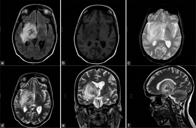
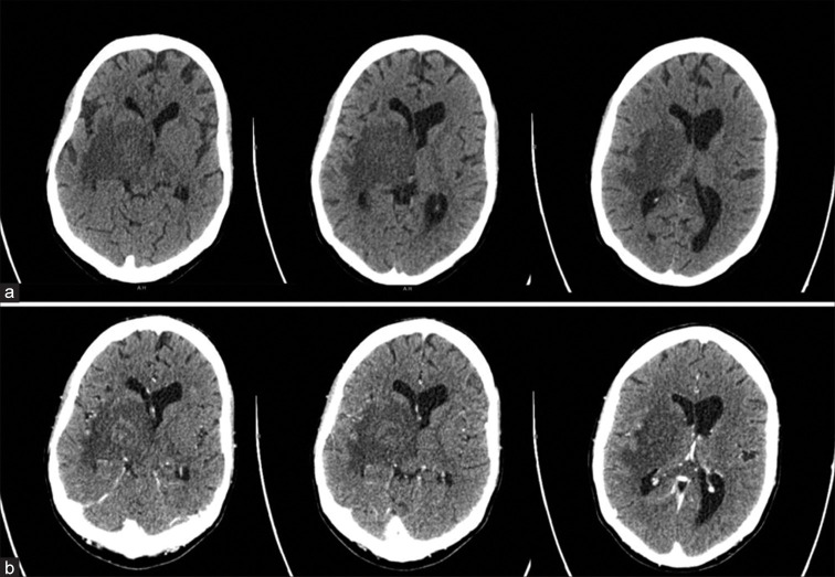
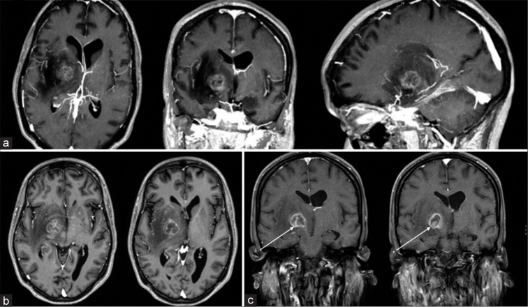
Background: Cerebral toxoplasmosis is a relatively rare disorder that usually affects immunocompromised patients. The most common scenario occurs among human immunodeficiency virus (HIV)-positive patients. In those patients, toxoplasmosis is the most frequent cause of expansive brain lesion and continues to cause elevated morbidity and mortality. In typical cases of toxoplasmosis, both computed tomography and magnetic resonance imaging reveal single/ multiple nodular or ring-enhancing lesions with surrounding edema. Nevertheless, cases of cerebral toxoplasmosis with atypical radiological features have been reported. Diagnosis can be obtained by finding organisms in the cerebrospinal fluid or in stereotactic biopsy samples of the brain lesion. If untreated, cerebral toxoplasmosis is uniformly fatal, so prompt diagnosis is mandatory. A prompt diagnosis is necessary, as untreated cerebral toxoplasmosis is uniformly fatal. Case Description: We discuss imaging and clinical findings of a patient – not aware of being HIV-positive – with a solitary atypical brain localization of toxoplasmosis mimicking a brain tumor. Conclusion: Although relatively uncommon, neurosurgeons should be aware of the potential occurrence of cerebral toxoplasmosis. High index of suspicion is needed for timely diagnosis and prompt initiation of therapy.


 分享
分享
 求助内容:
求助内容: 应助结果提醒方式:
 扫码关注我们
