{"title":"胰腺外植体培养具有间充质分化潜能的骨髓单核细胞样祖细胞的鉴定。","authors":"Marc-Estienne Roehrich, Giuseppe Vassalli","doi":"10.1155/2012/429868","DOIUrl":null,"url":null,"abstract":"<p><p>Progenitor cells can be obtained by outgrowth from tissue explants during primary ex vivo tissue culture. We have isolated and characterized cells outgrown from neonatal mouse pancreatic explants. A relatively uniform population of cells showing a distinctive morphology emerged over time in culture. This population expressed monocyte/macrophage and hematopoietic markers (CD11b(+) and CD45(+)), and some stromal-related markers (CD44(+) and CD29(+)), but not mesenchymal stem cell (MSC)-defining markers (CD90(-) and CD105(-)) nor endothelial (CD31(-)) or stem cell-associated markers (CD133(-) and stem cell antigen-1; Sca-1(-)). Cells could be maintained in culture as a plastic-adherent monolayer in culture medium (MesenCult MSC) for more than 1 year. Cells spontaneously formed sphere clusters \"pancreatospheres\" which, however, were nonclonal. When cultured in appropriate media, cells differentiated into multiple mesenchymal lineages (fat, cartilage, and bone). Positive dithizone staining suggested that a subset of cells differentiated into insulin-producing cells. However, further studies are needed to characterize the endocrine potential of these cells. These findings indicate that a myelomonocytoid population from pancreatic explant outgrowths has mesenchymal differentiation potential. These results are in line with recent data onmonocyte-derivedmesenchymal progenitors (MOMPs).</p>","PeriodicalId":9268,"journal":{"name":"Biotechnology Research International","volume":"2012 ","pages":"429868"},"PeriodicalIF":0.0000,"publicationDate":"2012-01-01","publicationTypes":"Journal Article","fieldsOfStudy":null,"isOpenAccess":false,"openAccessPdf":"https://www.ncbi.nlm.nih.gov/pmc/articles/PMC3431127/pdf/","citationCount":"2","resultStr":"{\"title\":\"Characterization of myelomonocytoid progenitor cells with mesenchymal differentiation potential obtained by outgrowth from pancreas explants.\",\"authors\":\"Marc-Estienne Roehrich, Giuseppe Vassalli\",\"doi\":\"10.1155/2012/429868\",\"DOIUrl\":null,\"url\":null,\"abstract\":\"<p><p>Progenitor cells can be obtained by outgrowth from tissue explants during primary ex vivo tissue culture. We have isolated and characterized cells outgrown from neonatal mouse pancreatic explants. A relatively uniform population of cells showing a distinctive morphology emerged over time in culture. This population expressed monocyte/macrophage and hematopoietic markers (CD11b(+) and CD45(+)), and some stromal-related markers (CD44(+) and CD29(+)), but not mesenchymal stem cell (MSC)-defining markers (CD90(-) and CD105(-)) nor endothelial (CD31(-)) or stem cell-associated markers (CD133(-) and stem cell antigen-1; Sca-1(-)). Cells could be maintained in culture as a plastic-adherent monolayer in culture medium (MesenCult MSC) for more than 1 year. Cells spontaneously formed sphere clusters \\\"pancreatospheres\\\" which, however, were nonclonal. When cultured in appropriate media, cells differentiated into multiple mesenchymal lineages (fat, cartilage, and bone). Positive dithizone staining suggested that a subset of cells differentiated into insulin-producing cells. However, further studies are needed to characterize the endocrine potential of these cells. These findings indicate that a myelomonocytoid population from pancreatic explant outgrowths has mesenchymal differentiation potential. These results are in line with recent data onmonocyte-derivedmesenchymal progenitors (MOMPs).</p>\",\"PeriodicalId\":9268,\"journal\":{\"name\":\"Biotechnology Research International\",\"volume\":\"2012 \",\"pages\":\"429868\"},\"PeriodicalIF\":0.0000,\"publicationDate\":\"2012-01-01\",\"publicationTypes\":\"Journal Article\",\"fieldsOfStudy\":null,\"isOpenAccess\":false,\"openAccessPdf\":\"https://www.ncbi.nlm.nih.gov/pmc/articles/PMC3431127/pdf/\",\"citationCount\":\"2\",\"resultStr\":null,\"platform\":\"Semanticscholar\",\"paperid\":null,\"PeriodicalName\":\"Biotechnology Research International\",\"FirstCategoryId\":\"1085\",\"ListUrlMain\":\"https://doi.org/10.1155/2012/429868\",\"RegionNum\":0,\"RegionCategory\":null,\"ArticlePicture\":[],\"TitleCN\":null,\"AbstractTextCN\":null,\"PMCID\":null,\"EPubDate\":\"\",\"PubModel\":\"\",\"JCR\":\"\",\"JCRName\":\"\",\"Score\":null,\"Total\":0}","platform":"Semanticscholar","paperid":null,"PeriodicalName":"Biotechnology Research International","FirstCategoryId":"1085","ListUrlMain":"https://doi.org/10.1155/2012/429868","RegionNum":0,"RegionCategory":null,"ArticlePicture":[],"TitleCN":null,"AbstractTextCN":null,"PMCID":null,"EPubDate":"","PubModel":"","JCR":"","JCRName":"","Score":null,"Total":0}
Characterization of myelomonocytoid progenitor cells with mesenchymal differentiation potential obtained by outgrowth from pancreas explants.
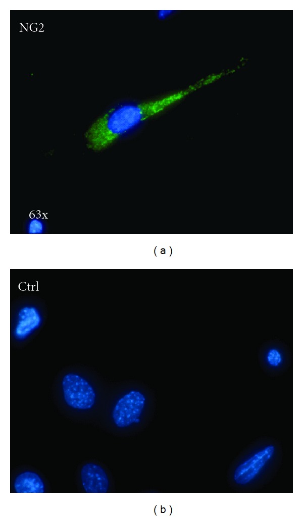
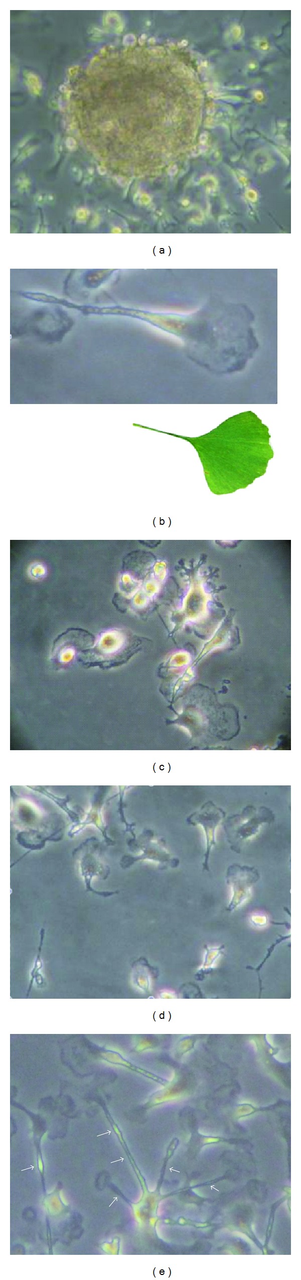
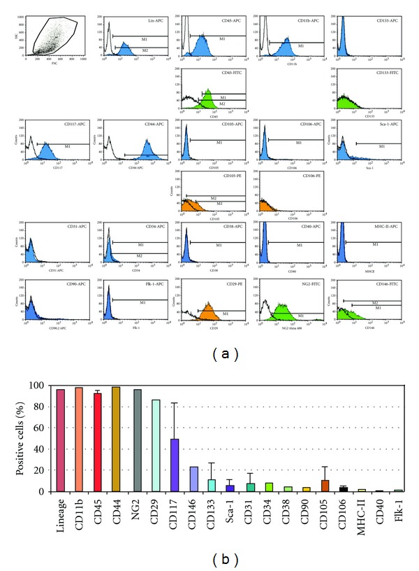
Progenitor cells can be obtained by outgrowth from tissue explants during primary ex vivo tissue culture. We have isolated and characterized cells outgrown from neonatal mouse pancreatic explants. A relatively uniform population of cells showing a distinctive morphology emerged over time in culture. This population expressed monocyte/macrophage and hematopoietic markers (CD11b(+) and CD45(+)), and some stromal-related markers (CD44(+) and CD29(+)), but not mesenchymal stem cell (MSC)-defining markers (CD90(-) and CD105(-)) nor endothelial (CD31(-)) or stem cell-associated markers (CD133(-) and stem cell antigen-1; Sca-1(-)). Cells could be maintained in culture as a plastic-adherent monolayer in culture medium (MesenCult MSC) for more than 1 year. Cells spontaneously formed sphere clusters "pancreatospheres" which, however, were nonclonal. When cultured in appropriate media, cells differentiated into multiple mesenchymal lineages (fat, cartilage, and bone). Positive dithizone staining suggested that a subset of cells differentiated into insulin-producing cells. However, further studies are needed to characterize the endocrine potential of these cells. These findings indicate that a myelomonocytoid population from pancreatic explant outgrowths has mesenchymal differentiation potential. These results are in line with recent data onmonocyte-derivedmesenchymal progenitors (MOMPs).


 分享
分享

 求助内容:
求助内容: 应助结果提醒方式:
 扫码关注我们
