{"title":"下颌支内侧凹陷的特征:不同矢状面骨型的CBCT分析。","authors":"Mahvash Hasani, Maryam Karandish, Yalda Salari","doi":"10.30476/dentjods.2022.89659.1427","DOIUrl":null,"url":null,"abstract":"<p><strong>Statement of the problem: </strong>Medial depression of the mandibular ramus (MDMR) as a normal anatomical variation might complicate orthognatic surgeries that involve ramus. When planning an orthognatic surgery, it is clinically valuable to notice MDMR in osteotomy site to decrease the risk of failure.</p><p><strong>Purpose: </strong>The aim of present study was to evaluate the prevalence as well as characteristics of MDMR in three skeletal sagittal classifications.</p><p><strong>Materials and method: </strong>This cross sectional study evaluated 530 cone beam computed tomography (CBCT) scans, of which 220 were enrolled. The skeletal sagittal classification, the presence of MDMR, the shape, depth, and width of MDMR were recorded for each patient by two examiners. Chi-square test was performed to determine the differences between three skeletal sagittal groups and between two genders.</p><p><strong>Results: </strong>The overall prevalence of MDMR was 60.45%. MDMR was mostly detected in class III (76.92%), followed by class II (76.66%), and class I (54.87%). In the analyzed CBCT scans, semi-lunar was the most common shape detected (42.85%), followed by triangular (30.82%), circular (18.04%), and tear-drop (8.27%). The depth of MDMR was not significantly different between three sagittal groups and between genders; however, the width of MDMR was higher in class III group and in male patients. In the present study, MDMR was found to be more common in patients with class II and class III skeletal classifications. Although, MDMR was more frequent in class III, the difference between class II and class III was not significant.</p><p><strong>Conclusion: </strong>More caution is needed during orthognatic surgery in patients with dentoskeletal deformities during the splitting of the ramus. Moreover, higher width of MDMR in class III and male patients should be concerned when planning an orthognatic surgery for these patients.</p>","PeriodicalId":73702,"journal":{"name":"Journal of dentistry (Shiraz, Iran)","volume":"24 1","pages":"1-6"},"PeriodicalIF":0.0000,"publicationDate":"2023-03-01","publicationTypes":"Journal Article","fieldsOfStudy":null,"isOpenAccess":false,"openAccessPdf":"https://www.ncbi.nlm.nih.gov/pmc/articles/PMC9971603/pdf/","citationCount":"0","resultStr":"{\"title\":\"Characteristics of Medial Depression of the Mandibular Ramus: A CBCT Analysis in Different Sagittal Skeletal Patterns.\",\"authors\":\"Mahvash Hasani, Maryam Karandish, Yalda Salari\",\"doi\":\"10.30476/dentjods.2022.89659.1427\",\"DOIUrl\":null,\"url\":null,\"abstract\":\"<p><strong>Statement of the problem: </strong>Medial depression of the mandibular ramus (MDMR) as a normal anatomical variation might complicate orthognatic surgeries that involve ramus. When planning an orthognatic surgery, it is clinically valuable to notice MDMR in osteotomy site to decrease the risk of failure.</p><p><strong>Purpose: </strong>The aim of present study was to evaluate the prevalence as well as characteristics of MDMR in three skeletal sagittal classifications.</p><p><strong>Materials and method: </strong>This cross sectional study evaluated 530 cone beam computed tomography (CBCT) scans, of which 220 were enrolled. The skeletal sagittal classification, the presence of MDMR, the shape, depth, and width of MDMR were recorded for each patient by two examiners. Chi-square test was performed to determine the differences between three skeletal sagittal groups and between two genders.</p><p><strong>Results: </strong>The overall prevalence of MDMR was 60.45%. MDMR was mostly detected in class III (76.92%), followed by class II (76.66%), and class I (54.87%). In the analyzed CBCT scans, semi-lunar was the most common shape detected (42.85%), followed by triangular (30.82%), circular (18.04%), and tear-drop (8.27%). The depth of MDMR was not significantly different between three sagittal groups and between genders; however, the width of MDMR was higher in class III group and in male patients. In the present study, MDMR was found to be more common in patients with class II and class III skeletal classifications. Although, MDMR was more frequent in class III, the difference between class II and class III was not significant.</p><p><strong>Conclusion: </strong>More caution is needed during orthognatic surgery in patients with dentoskeletal deformities during the splitting of the ramus. Moreover, higher width of MDMR in class III and male patients should be concerned when planning an orthognatic surgery for these patients.</p>\",\"PeriodicalId\":73702,\"journal\":{\"name\":\"Journal of dentistry (Shiraz, Iran)\",\"volume\":\"24 1\",\"pages\":\"1-6\"},\"PeriodicalIF\":0.0000,\"publicationDate\":\"2023-03-01\",\"publicationTypes\":\"Journal Article\",\"fieldsOfStudy\":null,\"isOpenAccess\":false,\"openAccessPdf\":\"https://www.ncbi.nlm.nih.gov/pmc/articles/PMC9971603/pdf/\",\"citationCount\":\"0\",\"resultStr\":null,\"platform\":\"Semanticscholar\",\"paperid\":null,\"PeriodicalName\":\"Journal of dentistry (Shiraz, Iran)\",\"FirstCategoryId\":\"1085\",\"ListUrlMain\":\"https://doi.org/10.30476/dentjods.2022.89659.1427\",\"RegionNum\":0,\"RegionCategory\":null,\"ArticlePicture\":[],\"TitleCN\":null,\"AbstractTextCN\":null,\"PMCID\":null,\"EPubDate\":\"\",\"PubModel\":\"\",\"JCR\":\"\",\"JCRName\":\"\",\"Score\":null,\"Total\":0}","platform":"Semanticscholar","paperid":null,"PeriodicalName":"Journal of dentistry (Shiraz, Iran)","FirstCategoryId":"1085","ListUrlMain":"https://doi.org/10.30476/dentjods.2022.89659.1427","RegionNum":0,"RegionCategory":null,"ArticlePicture":[],"TitleCN":null,"AbstractTextCN":null,"PMCID":null,"EPubDate":"","PubModel":"","JCR":"","JCRName":"","Score":null,"Total":0}
Characteristics of Medial Depression of the Mandibular Ramus: A CBCT Analysis in Different Sagittal Skeletal Patterns.
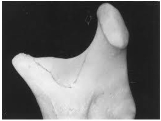
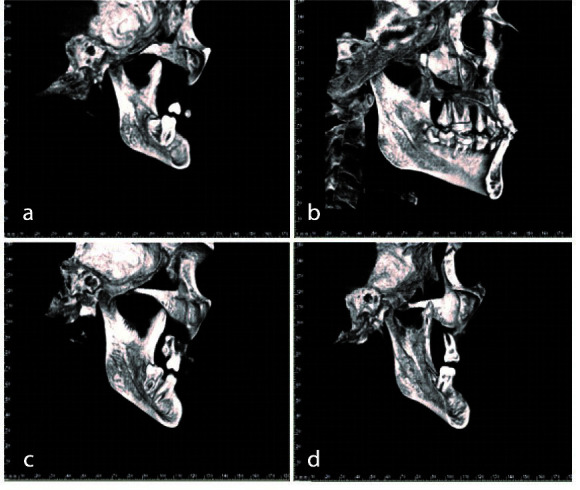
Statement of the problem: Medial depression of the mandibular ramus (MDMR) as a normal anatomical variation might complicate orthognatic surgeries that involve ramus. When planning an orthognatic surgery, it is clinically valuable to notice MDMR in osteotomy site to decrease the risk of failure.
Purpose: The aim of present study was to evaluate the prevalence as well as characteristics of MDMR in three skeletal sagittal classifications.
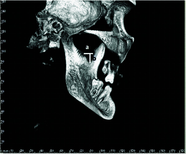
Materials and method: This cross sectional study evaluated 530 cone beam computed tomography (CBCT) scans, of which 220 were enrolled. The skeletal sagittal classification, the presence of MDMR, the shape, depth, and width of MDMR were recorded for each patient by two examiners. Chi-square test was performed to determine the differences between three skeletal sagittal groups and between two genders.
Results: The overall prevalence of MDMR was 60.45%. MDMR was mostly detected in class III (76.92%), followed by class II (76.66%), and class I (54.87%). In the analyzed CBCT scans, semi-lunar was the most common shape detected (42.85%), followed by triangular (30.82%), circular (18.04%), and tear-drop (8.27%). The depth of MDMR was not significantly different between three sagittal groups and between genders; however, the width of MDMR was higher in class III group and in male patients. In the present study, MDMR was found to be more common in patients with class II and class III skeletal classifications. Although, MDMR was more frequent in class III, the difference between class II and class III was not significant.
Conclusion: More caution is needed during orthognatic surgery in patients with dentoskeletal deformities during the splitting of the ramus. Moreover, higher width of MDMR in class III and male patients should be concerned when planning an orthognatic surgery for these patients.


 分享
分享

 求助内容:
求助内容: 应助结果提醒方式:
 扫码关注我们
