Scherwin Mahmoudi, Marvin Lange, Lukas Lenga, Ibrahim Yel, Vitali Koch, Christian Booz, Simon Martin, Simon Bernatz, Thomas Vogl, Moritz Albrecht, Jan-Erik Scholtz
{"title":"利用噪声优化的虚拟单能图像重建挽救低对比腹部CT研究。","authors":"Scherwin Mahmoudi, Marvin Lange, Lukas Lenga, Ibrahim Yel, Vitali Koch, Christian Booz, Simon Martin, Simon Bernatz, Thomas Vogl, Moritz Albrecht, Jan-Erik Scholtz","doi":"10.1259/bjro.20220006","DOIUrl":null,"url":null,"abstract":"<p><strong>Objectives: </strong>To assess the impact of noise-optimised virtual monoenergetic imaging (VMI+) on image quality and diagnostic evaluation in abdominal dual-energy CT scans with impaired portal-venous contrast.</p><p><strong>Methods: </strong>We screened 11,746 patients who underwent portal-venous abdominal dual-energy CT for cancer staging between 08/2014 and 11/2019 and identified those with poor portal-venous contrast.Standard linearly-blended image series and VMI+ image series at 40, 50, and 60 keV were reconstructed. Signal-to-noise ratio (SNR) and contrast-to-noise ratio (CNR) of abdominal organs and vascular structures were calculated. Image noise, image contrast and overall image quality were rated by three radiologists using 5-point Likert scale.</p><p><strong>Results: </strong>452 of 11,746 (4%) exams were poorly opacified. We excluded 190 cases due to incomplete datasets or multiple exams of the same patient with a final study group of 262. Highest CNR values in all abdominal organs (liver, 6.4 ± 3.0; kidney, 17.4 ± 7.5; spleen, 8.0 ± 3.5) and vascular structures (aorta, 16.0 ± 7.3; intrahepatic vein, 11.3 ± 4.7; portal vein, 15.5 ± 6.7) were measured at 40 keV VMI+ with significantly superior values compared to all other series. In subjective analysis, highest image contrast was seen at 40 keV VMI+ (4.8 ± 0.4), whereas overall image quality peaked at 50 keV VMI+ (4.2 ± 0.5) with significantly superior results compared to all other series (<i>p</i> < 0.001).</p><p><strong>Conclusions: </strong>Image reconstruction using VMI+ algorithm at 50 keV significantly improves image contrast and image quality of originally poorly opacified abdominal CT scans and reduces the number of non-diagnostic scans.</p><p><strong>Advances in knowledge: </strong>We validated the impact of VMI+ reconstructions in poorly attenuated DECT studies of the abdomen in a big data cohort.</p>","PeriodicalId":72419,"journal":{"name":"BJR open","volume":"4 1","pages":"20220006"},"PeriodicalIF":2.1000,"publicationDate":"2022-01-01","publicationTypes":"Journal Article","fieldsOfStudy":null,"isOpenAccess":false,"openAccessPdf":"https://www.ncbi.nlm.nih.gov/pmc/articles/PMC9446156/pdf/","citationCount":"1","resultStr":"{\"title\":\"Salvaging low contrast abdominal CT studies using noise-optimised virtual monoenergetic image reconstruction.\",\"authors\":\"Scherwin Mahmoudi, Marvin Lange, Lukas Lenga, Ibrahim Yel, Vitali Koch, Christian Booz, Simon Martin, Simon Bernatz, Thomas Vogl, Moritz Albrecht, Jan-Erik Scholtz\",\"doi\":\"10.1259/bjro.20220006\",\"DOIUrl\":null,\"url\":null,\"abstract\":\"<p><strong>Objectives: </strong>To assess the impact of noise-optimised virtual monoenergetic imaging (VMI+) on image quality and diagnostic evaluation in abdominal dual-energy CT scans with impaired portal-venous contrast.</p><p><strong>Methods: </strong>We screened 11,746 patients who underwent portal-venous abdominal dual-energy CT for cancer staging between 08/2014 and 11/2019 and identified those with poor portal-venous contrast.Standard linearly-blended image series and VMI+ image series at 40, 50, and 60 keV were reconstructed. Signal-to-noise ratio (SNR) and contrast-to-noise ratio (CNR) of abdominal organs and vascular structures were calculated. Image noise, image contrast and overall image quality were rated by three radiologists using 5-point Likert scale.</p><p><strong>Results: </strong>452 of 11,746 (4%) exams were poorly opacified. We excluded 190 cases due to incomplete datasets or multiple exams of the same patient with a final study group of 262. Highest CNR values in all abdominal organs (liver, 6.4 ± 3.0; kidney, 17.4 ± 7.5; spleen, 8.0 ± 3.5) and vascular structures (aorta, 16.0 ± 7.3; intrahepatic vein, 11.3 ± 4.7; portal vein, 15.5 ± 6.7) were measured at 40 keV VMI+ with significantly superior values compared to all other series. In subjective analysis, highest image contrast was seen at 40 keV VMI+ (4.8 ± 0.4), whereas overall image quality peaked at 50 keV VMI+ (4.2 ± 0.5) with significantly superior results compared to all other series (<i>p</i> < 0.001).</p><p><strong>Conclusions: </strong>Image reconstruction using VMI+ algorithm at 50 keV significantly improves image contrast and image quality of originally poorly opacified abdominal CT scans and reduces the number of non-diagnostic scans.</p><p><strong>Advances in knowledge: </strong>We validated the impact of VMI+ reconstructions in poorly attenuated DECT studies of the abdomen in a big data cohort.</p>\",\"PeriodicalId\":72419,\"journal\":{\"name\":\"BJR open\",\"volume\":\"4 1\",\"pages\":\"20220006\"},\"PeriodicalIF\":2.1000,\"publicationDate\":\"2022-01-01\",\"publicationTypes\":\"Journal Article\",\"fieldsOfStudy\":null,\"isOpenAccess\":false,\"openAccessPdf\":\"https://www.ncbi.nlm.nih.gov/pmc/articles/PMC9446156/pdf/\",\"citationCount\":\"1\",\"resultStr\":null,\"platform\":\"Semanticscholar\",\"paperid\":null,\"PeriodicalName\":\"BJR open\",\"FirstCategoryId\":\"1085\",\"ListUrlMain\":\"https://doi.org/10.1259/bjro.20220006\",\"RegionNum\":0,\"RegionCategory\":null,\"ArticlePicture\":[],\"TitleCN\":null,\"AbstractTextCN\":null,\"PMCID\":null,\"EPubDate\":\"\",\"PubModel\":\"\",\"JCR\":\"\",\"JCRName\":\"\",\"Score\":null,\"Total\":0}","platform":"Semanticscholar","paperid":null,"PeriodicalName":"BJR open","FirstCategoryId":"1085","ListUrlMain":"https://doi.org/10.1259/bjro.20220006","RegionNum":0,"RegionCategory":null,"ArticlePicture":[],"TitleCN":null,"AbstractTextCN":null,"PMCID":null,"EPubDate":"","PubModel":"","JCR":"","JCRName":"","Score":null,"Total":0}
Objectives: To assess the impact of noise-optimised virtual monoenergetic imaging (VMI+) on image quality and diagnostic evaluation in abdominal dual-energy CT scans with impaired portal-venous contrast.
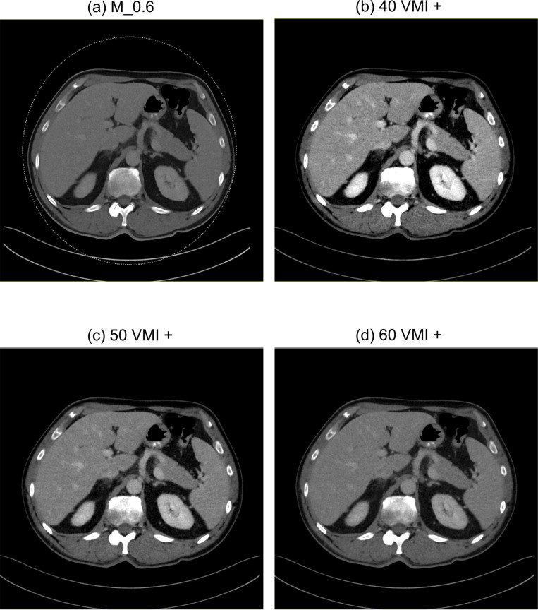
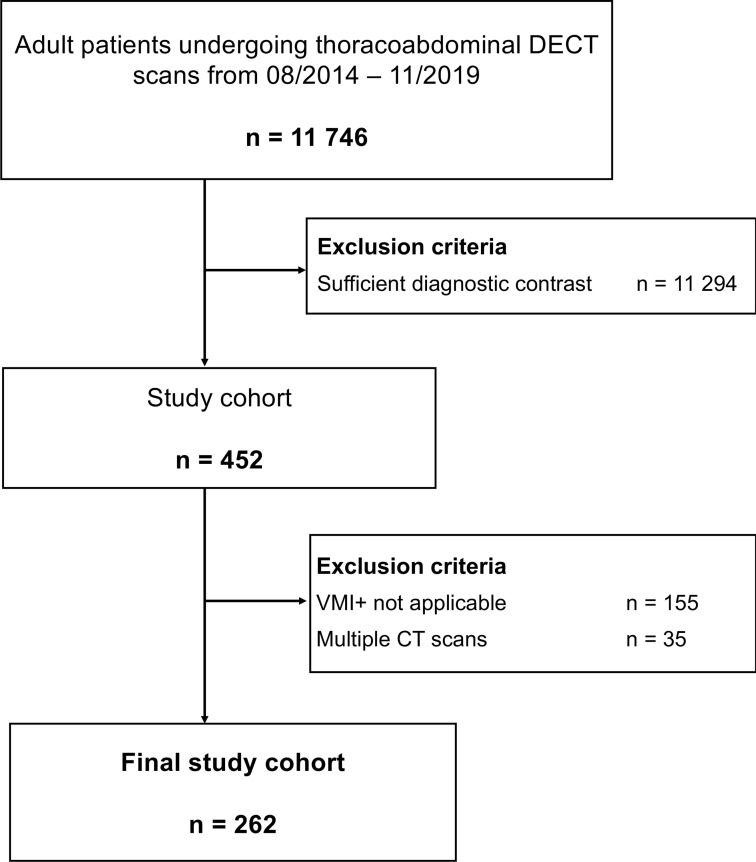
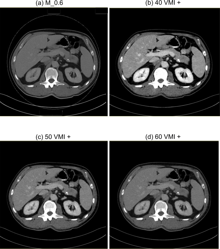
Methods: We screened 11,746 patients who underwent portal-venous abdominal dual-energy CT for cancer staging between 08/2014 and 11/2019 and identified those with poor portal-venous contrast.Standard linearly-blended image series and VMI+ image series at 40, 50, and 60 keV were reconstructed. Signal-to-noise ratio (SNR) and contrast-to-noise ratio (CNR) of abdominal organs and vascular structures were calculated. Image noise, image contrast and overall image quality were rated by three radiologists using 5-point Likert scale.
Results: 452 of 11,746 (4%) exams were poorly opacified. We excluded 190 cases due to incomplete datasets or multiple exams of the same patient with a final study group of 262. Highest CNR values in all abdominal organs (liver, 6.4 ± 3.0; kidney, 17.4 ± 7.5; spleen, 8.0 ± 3.5) and vascular structures (aorta, 16.0 ± 7.3; intrahepatic vein, 11.3 ± 4.7; portal vein, 15.5 ± 6.7) were measured at 40 keV VMI+ with significantly superior values compared to all other series. In subjective analysis, highest image contrast was seen at 40 keV VMI+ (4.8 ± 0.4), whereas overall image quality peaked at 50 keV VMI+ (4.2 ± 0.5) with significantly superior results compared to all other series (p < 0.001).
Conclusions: Image reconstruction using VMI+ algorithm at 50 keV significantly improves image contrast and image quality of originally poorly opacified abdominal CT scans and reduces the number of non-diagnostic scans.
Advances in knowledge: We validated the impact of VMI+ reconstructions in poorly attenuated DECT studies of the abdomen in a big data cohort.


 分享
分享

 求助内容:
求助内容: 应助结果提醒方式:
 扫码关注我们
