Waleed Althobaity, Ayman Aldeheshi, Mnahi Bin Saeedan
{"title":"原发性胸壁包虫病:1例多模态影像学表现。","authors":"Waleed Althobaity, Ayman Aldeheshi, Mnahi Bin Saeedan","doi":"10.1155/2023/5313067","DOIUrl":null,"url":null,"abstract":"<p><p>Primary chest wall hydatid cyst is a very rare disease in endemic areas. This case report describes a 22-year-old male patient with a 3-year history of chronic left chest pain. He had a history of close animal contact in childhood. Chest computed tomography (CT) scan showed a left upper posterior paravertebral cystic mass with peripheral and intrinsic calcifications. Fluorine-18 fluorodeoxyglucose (F-18 FDG) positron emission tomography (PET) scan showed no significant FDG uptake. Magnetic resonance imaging (MRI) showed a left paravertebral cystic mass with daughter cysts and a peripheral low T2 wall, compatible with hydatid disease. Medical treatment was started, and a follow-up MRI showed rupture of hydatid cysts. The patient underwent surgical resection, and a hydatid disease diagnosis was confirmed by histopathologic examination. During the postoperative hospital course, the patient developed pneumothorax which was successfully treated with a surgical procedure. The patient was discharged with medical treatment (albendazole). In conclusion, this case highlights the importance of considering hydatid disease in the differential diagnosis of chest wall cystic masses, especially in endemic regions, and the value of multimodality imaging in diagnosis and treatment planning.</p>","PeriodicalId":30326,"journal":{"name":"Case Reports in Radiology","volume":"2023 ","pages":"5313067"},"PeriodicalIF":0.0000,"publicationDate":"2023-01-01","publicationTypes":"Journal Article","fieldsOfStudy":null,"isOpenAccess":false,"openAccessPdf":"https://www.ncbi.nlm.nih.gov/pmc/articles/PMC10118874/pdf/","citationCount":"0","resultStr":"{\"title\":\"Primary Chest Wall Hydatid Disease: A Case Report with Multimodality Imaging Findings.\",\"authors\":\"Waleed Althobaity, Ayman Aldeheshi, Mnahi Bin Saeedan\",\"doi\":\"10.1155/2023/5313067\",\"DOIUrl\":null,\"url\":null,\"abstract\":\"<p><p>Primary chest wall hydatid cyst is a very rare disease in endemic areas. This case report describes a 22-year-old male patient with a 3-year history of chronic left chest pain. He had a history of close animal contact in childhood. Chest computed tomography (CT) scan showed a left upper posterior paravertebral cystic mass with peripheral and intrinsic calcifications. Fluorine-18 fluorodeoxyglucose (F-18 FDG) positron emission tomography (PET) scan showed no significant FDG uptake. Magnetic resonance imaging (MRI) showed a left paravertebral cystic mass with daughter cysts and a peripheral low T2 wall, compatible with hydatid disease. Medical treatment was started, and a follow-up MRI showed rupture of hydatid cysts. The patient underwent surgical resection, and a hydatid disease diagnosis was confirmed by histopathologic examination. During the postoperative hospital course, the patient developed pneumothorax which was successfully treated with a surgical procedure. The patient was discharged with medical treatment (albendazole). In conclusion, this case highlights the importance of considering hydatid disease in the differential diagnosis of chest wall cystic masses, especially in endemic regions, and the value of multimodality imaging in diagnosis and treatment planning.</p>\",\"PeriodicalId\":30326,\"journal\":{\"name\":\"Case Reports in Radiology\",\"volume\":\"2023 \",\"pages\":\"5313067\"},\"PeriodicalIF\":0.0000,\"publicationDate\":\"2023-01-01\",\"publicationTypes\":\"Journal Article\",\"fieldsOfStudy\":null,\"isOpenAccess\":false,\"openAccessPdf\":\"https://www.ncbi.nlm.nih.gov/pmc/articles/PMC10118874/pdf/\",\"citationCount\":\"0\",\"resultStr\":null,\"platform\":\"Semanticscholar\",\"paperid\":null,\"PeriodicalName\":\"Case Reports in Radiology\",\"FirstCategoryId\":\"1085\",\"ListUrlMain\":\"https://doi.org/10.1155/2023/5313067\",\"RegionNum\":0,\"RegionCategory\":null,\"ArticlePicture\":[],\"TitleCN\":null,\"AbstractTextCN\":null,\"PMCID\":null,\"EPubDate\":\"\",\"PubModel\":\"\",\"JCR\":\"\",\"JCRName\":\"\",\"Score\":null,\"Total\":0}","platform":"Semanticscholar","paperid":null,"PeriodicalName":"Case Reports in Radiology","FirstCategoryId":"1085","ListUrlMain":"https://doi.org/10.1155/2023/5313067","RegionNum":0,"RegionCategory":null,"ArticlePicture":[],"TitleCN":null,"AbstractTextCN":null,"PMCID":null,"EPubDate":"","PubModel":"","JCR":"","JCRName":"","Score":null,"Total":0}
Primary Chest Wall Hydatid Disease: A Case Report with Multimodality Imaging Findings.
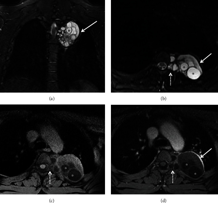
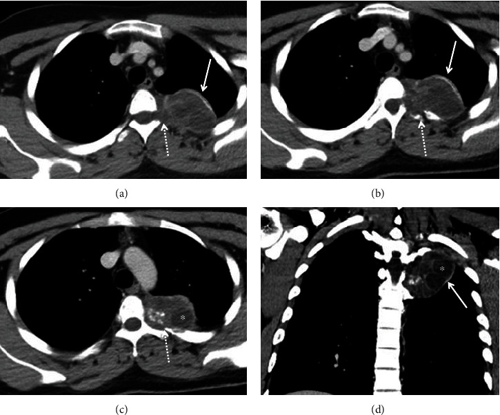
Primary chest wall hydatid cyst is a very rare disease in endemic areas. This case report describes a 22-year-old male patient with a 3-year history of chronic left chest pain. He had a history of close animal contact in childhood. Chest computed tomography (CT) scan showed a left upper posterior paravertebral cystic mass with peripheral and intrinsic calcifications. Fluorine-18 fluorodeoxyglucose (F-18 FDG) positron emission tomography (PET) scan showed no significant FDG uptake. Magnetic resonance imaging (MRI) showed a left paravertebral cystic mass with daughter cysts and a peripheral low T2 wall, compatible with hydatid disease. Medical treatment was started, and a follow-up MRI showed rupture of hydatid cysts. The patient underwent surgical resection, and a hydatid disease diagnosis was confirmed by histopathologic examination. During the postoperative hospital course, the patient developed pneumothorax which was successfully treated with a surgical procedure. The patient was discharged with medical treatment (albendazole). In conclusion, this case highlights the importance of considering hydatid disease in the differential diagnosis of chest wall cystic masses, especially in endemic regions, and the value of multimodality imaging in diagnosis and treatment planning.


 分享
分享

 求助内容:
求助内容: 应助结果提醒方式:
 扫码关注我们
