Matteo Bonatti, Nicolò De Pretis, Giulia A Zamboni, Alessandro Brillo, Stefano Francesco Crinò, Riccardo Valletta, Fabio Lombardo, Giancarlo Mansueto, Luca Frulloni
{"title":"十二指肠旁胰腺炎的影像学:系统回顾。","authors":"Matteo Bonatti, Nicolò De Pretis, Giulia A Zamboni, Alessandro Brillo, Stefano Francesco Crinò, Riccardo Valletta, Fabio Lombardo, Giancarlo Mansueto, Luca Frulloni","doi":"10.4329/wjr.v15.i2.42","DOIUrl":null,"url":null,"abstract":"<p><strong>Background: </strong>Paraduodenal pancreatitis (PP) represents a diagnostic challenge, especially in non-referral centers, given its potential imaging overlap with pancreatic cancer. There are two main histological variants of PP, the cystic and the solid, with slightly different imaging appearances. Moreover, imaging findings in PP may change over time because of disease progression and/or as an effect of its risk factors exposition, namely alcohol intake and smoking.</p><p><strong>Aim: </strong>To describe multimodality imaging findings in patients affected by PP to help clinicians in the differential diagnosis with pancreatic cancer.</p><p><strong>Methods: </strong>The systematic review was conducted according to the Preferred Reporting Items for Systematic reviews and Meta-analyses 2009 guidelines. A Literature search was performed on PubMed, Embase and Cochrane Library using (groove pancreatitis [Title/Abstract]) OR (PP [Title/Abstract]) as key words. A total of 593 articles were considered for inclusion. After eliminating duplicates, and title and abstract screening, 53 full-text articles were assessed for eligibility. Eligibility criteria were: Original studies including 8 or more patients, fully written in English, describing imaging findings in PP, with pathological confirmation or clinical-radiological follow-up as the gold standard. Finally, 14 studies were included in our systematic review.</p><p><strong>Results: </strong>Computed tomography (CT) findings were described in 292 patients, magnetic resonance imaging (MRI) findings in 231 and endoscopic ultrasound (EUS) findings in 115. Duodenal wall thickening was observed in 88.8% of the cases: Detection rate was 96.5% at EUS, 91.0% at MRI and 84.1% at CT. Second duodenal portion increased enhancement was recognizable in 76.3% of the cases: Detection rate was 84.4% at MRI and 72.1% at CT. Cysts within the duodenal wall were detected in 82.6% of the cases: Detection rate was 94.4% at EUS, 81.9% at MRI and 75.7% at CT. A solid mass in the groove region was described in 40.9% of the cases; in 78.3% of the cases, it showed patchy enhancement in the portal venous phase, and in 100% appeared iso/hyperintense during delayed phase imaging. Only 3.6% of the lesions showed restricted diffusion. The prevalence of radiological signs of chronic obstructive pancreatitis, namely main pancreatic duct dilatation, pancreatic calcifications, and pancreatic cysts, was extremely variable in the different articles.</p><p><strong>Conclusion: </strong>PP has peculiar imaging findings. MRI is the best radiological imaging modality for diagnosing PP, but EUS is more accurate than MRI in depicting duodenal wall alterations.</p>","PeriodicalId":23819,"journal":{"name":"World journal of radiology","volume":"15 2","pages":"42-55"},"PeriodicalIF":1.5000,"publicationDate":"2023-02-28","publicationTypes":"Journal Article","fieldsOfStudy":null,"isOpenAccess":false,"openAccessPdf":"https://ftp.ncbi.nlm.nih.gov/pub/pmc/oa_pdf/9b/37/WJR-15-42.PMC9979191.pdf","citationCount":"0","resultStr":"{\"title\":\"Imaging of paraduodenal pancreatitis: A systematic review.\",\"authors\":\"Matteo Bonatti, Nicolò De Pretis, Giulia A Zamboni, Alessandro Brillo, Stefano Francesco Crinò, Riccardo Valletta, Fabio Lombardo, Giancarlo Mansueto, Luca Frulloni\",\"doi\":\"10.4329/wjr.v15.i2.42\",\"DOIUrl\":null,\"url\":null,\"abstract\":\"<p><strong>Background: </strong>Paraduodenal pancreatitis (PP) represents a diagnostic challenge, especially in non-referral centers, given its potential imaging overlap with pancreatic cancer. There are two main histological variants of PP, the cystic and the solid, with slightly different imaging appearances. Moreover, imaging findings in PP may change over time because of disease progression and/or as an effect of its risk factors exposition, namely alcohol intake and smoking.</p><p><strong>Aim: </strong>To describe multimodality imaging findings in patients affected by PP to help clinicians in the differential diagnosis with pancreatic cancer.</p><p><strong>Methods: </strong>The systematic review was conducted according to the Preferred Reporting Items for Systematic reviews and Meta-analyses 2009 guidelines. A Literature search was performed on PubMed, Embase and Cochrane Library using (groove pancreatitis [Title/Abstract]) OR (PP [Title/Abstract]) as key words. A total of 593 articles were considered for inclusion. After eliminating duplicates, and title and abstract screening, 53 full-text articles were assessed for eligibility. Eligibility criteria were: Original studies including 8 or more patients, fully written in English, describing imaging findings in PP, with pathological confirmation or clinical-radiological follow-up as the gold standard. Finally, 14 studies were included in our systematic review.</p><p><strong>Results: </strong>Computed tomography (CT) findings were described in 292 patients, magnetic resonance imaging (MRI) findings in 231 and endoscopic ultrasound (EUS) findings in 115. Duodenal wall thickening was observed in 88.8% of the cases: Detection rate was 96.5% at EUS, 91.0% at MRI and 84.1% at CT. Second duodenal portion increased enhancement was recognizable in 76.3% of the cases: Detection rate was 84.4% at MRI and 72.1% at CT. Cysts within the duodenal wall were detected in 82.6% of the cases: Detection rate was 94.4% at EUS, 81.9% at MRI and 75.7% at CT. A solid mass in the groove region was described in 40.9% of the cases; in 78.3% of the cases, it showed patchy enhancement in the portal venous phase, and in 100% appeared iso/hyperintense during delayed phase imaging. Only 3.6% of the lesions showed restricted diffusion. The prevalence of radiological signs of chronic obstructive pancreatitis, namely main pancreatic duct dilatation, pancreatic calcifications, and pancreatic cysts, was extremely variable in the different articles.</p><p><strong>Conclusion: </strong>PP has peculiar imaging findings. MRI is the best radiological imaging modality for diagnosing PP, but EUS is more accurate than MRI in depicting duodenal wall alterations.</p>\",\"PeriodicalId\":23819,\"journal\":{\"name\":\"World journal of radiology\",\"volume\":\"15 2\",\"pages\":\"42-55\"},\"PeriodicalIF\":1.5000,\"publicationDate\":\"2023-02-28\",\"publicationTypes\":\"Journal Article\",\"fieldsOfStudy\":null,\"isOpenAccess\":false,\"openAccessPdf\":\"https://ftp.ncbi.nlm.nih.gov/pub/pmc/oa_pdf/9b/37/WJR-15-42.PMC9979191.pdf\",\"citationCount\":\"0\",\"resultStr\":null,\"platform\":\"Semanticscholar\",\"paperid\":null,\"PeriodicalName\":\"World journal of radiology\",\"FirstCategoryId\":\"1085\",\"ListUrlMain\":\"https://doi.org/10.4329/wjr.v15.i2.42\",\"RegionNum\":0,\"RegionCategory\":null,\"ArticlePicture\":[],\"TitleCN\":null,\"AbstractTextCN\":null,\"PMCID\":null,\"EPubDate\":\"\",\"PubModel\":\"\",\"JCR\":\"Q3\",\"JCRName\":\"RADIOLOGY, NUCLEAR MEDICINE & MEDICAL IMAGING\",\"Score\":null,\"Total\":0}","platform":"Semanticscholar","paperid":null,"PeriodicalName":"World journal of radiology","FirstCategoryId":"1085","ListUrlMain":"https://doi.org/10.4329/wjr.v15.i2.42","RegionNum":0,"RegionCategory":null,"ArticlePicture":[],"TitleCN":null,"AbstractTextCN":null,"PMCID":null,"EPubDate":"","PubModel":"","JCR":"Q3","JCRName":"RADIOLOGY, NUCLEAR MEDICINE & MEDICAL IMAGING","Score":null,"Total":0}
Imaging of paraduodenal pancreatitis: A systematic review.
Background: Paraduodenal pancreatitis (PP) represents a diagnostic challenge, especially in non-referral centers, given its potential imaging overlap with pancreatic cancer. There are two main histological variants of PP, the cystic and the solid, with slightly different imaging appearances. Moreover, imaging findings in PP may change over time because of disease progression and/or as an effect of its risk factors exposition, namely alcohol intake and smoking.
Aim: To describe multimodality imaging findings in patients affected by PP to help clinicians in the differential diagnosis with pancreatic cancer.
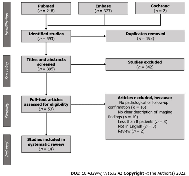
Methods: The systematic review was conducted according to the Preferred Reporting Items for Systematic reviews and Meta-analyses 2009 guidelines. A Literature search was performed on PubMed, Embase and Cochrane Library using (groove pancreatitis [Title/Abstract]) OR (PP [Title/Abstract]) as key words. A total of 593 articles were considered for inclusion. After eliminating duplicates, and title and abstract screening, 53 full-text articles were assessed for eligibility. Eligibility criteria were: Original studies including 8 or more patients, fully written in English, describing imaging findings in PP, with pathological confirmation or clinical-radiological follow-up as the gold standard. Finally, 14 studies were included in our systematic review.
Results: Computed tomography (CT) findings were described in 292 patients, magnetic resonance imaging (MRI) findings in 231 and endoscopic ultrasound (EUS) findings in 115. Duodenal wall thickening was observed in 88.8% of the cases: Detection rate was 96.5% at EUS, 91.0% at MRI and 84.1% at CT. Second duodenal portion increased enhancement was recognizable in 76.3% of the cases: Detection rate was 84.4% at MRI and 72.1% at CT. Cysts within the duodenal wall were detected in 82.6% of the cases: Detection rate was 94.4% at EUS, 81.9% at MRI and 75.7% at CT. A solid mass in the groove region was described in 40.9% of the cases; in 78.3% of the cases, it showed patchy enhancement in the portal venous phase, and in 100% appeared iso/hyperintense during delayed phase imaging. Only 3.6% of the lesions showed restricted diffusion. The prevalence of radiological signs of chronic obstructive pancreatitis, namely main pancreatic duct dilatation, pancreatic calcifications, and pancreatic cysts, was extremely variable in the different articles.
Conclusion: PP has peculiar imaging findings. MRI is the best radiological imaging modality for diagnosing PP, but EUS is more accurate than MRI in depicting duodenal wall alterations.


 分享
分享
 求助内容:
求助内容: 应助结果提醒方式:
 扫码关注我们
