{"title":"利用人工智能从3D图像中准确分割牙龈:动物试验研究。","authors":"Min Yang, Chenshuang Li, Wen Yang, Chider Chen, Chun-Hsi Chung, Nipul Tanna, Zhong Zheng","doi":"10.1186/s40510-023-00465-4","DOIUrl":null,"url":null,"abstract":"<p><strong>Background: </strong>Gingival phenotype plays an important role in dental diagnosis and treatment planning. Traditionally, determining the gingival phenotype is done by manual probing of the gingival soft tissues, an invasive and time-consuming procedure. This study aims to evaluate the feasibility and accuracy of an alternatively novel, non-invasive technology based on the precise 3-dimension (3D) soft tissue reconstruction from intraoral scanning and cone beam computed tomography (CBCT) to predict the gingival biotype.</p><p><strong>Methods: </strong>As a proof-of-concept, Yorkshire pig mandibles were scanned, and the CBCT data were fed into a deep-learning model to reconstruct the teeth and surrounding bone structure in 3D. By overlaying the CBCT scan with the intraoral scans, an accurate superposition was created and used for virtual measurements of the soft tissue thickness. Meanwhile, gingival thicknesses were also measured by a periodontal probe and digital caliper on the buccal and lingual sides at 3 mm apical to the gingival margin of the posterior teeth and compared with the virtual assessment at the same location. The data obtained from virtual and clinical measurements were compared by Wilcoxon matched-pairs signed-rank analysis, while their correlation was determined by Pearson's r value. The Mann-Whitney U test was used for intergroup comparisons of the amount of difference.</p><p><strong>Results: </strong>Among 108 investigated locations, the clinical and virtual measurements are strongly positively correlated (r = 0.9656, P < 0.0001), and only clinically insignificant differences (0.066 ± 0.223 mm) were observed between the two assessments. There is no difference in the agreement between the virtual and clinical measurements on sexually matured samples (0.087 ± 0.240 mm) and pre-pubertal samples (0.033 ± 0.195 mm). Noticeably, there is a greater agreement between the virtual and clinical measurements at the buccal sites (0.019 ± 0.233 mm) than at the lingual sites (0.116 ± 0.215 mm).</p><p><strong>Conclusion: </strong>In summary, the artificial intelligence-based virtual measurement proposed in this work provides an innovative technique potentially for accurately measuring soft tissue thickness using clinical routine 3D imaging systems, which will aid clinicians in generating a more comprehensive diagnosis with less invasive procedures and, in turn, optimize the treatment plans with more predictable outcomes.</p>","PeriodicalId":56071,"journal":{"name":"Progress in Orthodontics","volume":"24 1","pages":"14"},"PeriodicalIF":5.0000,"publicationDate":"2023-05-01","publicationTypes":"Journal Article","fieldsOfStudy":null,"isOpenAccess":false,"openAccessPdf":"https://www.ncbi.nlm.nih.gov/pmc/articles/PMC10149545/pdf/","citationCount":"1","resultStr":"{\"title\":\"Accurate gingival segmentation from 3D images with artificial intelligence: an animal pilot study.\",\"authors\":\"Min Yang, Chenshuang Li, Wen Yang, Chider Chen, Chun-Hsi Chung, Nipul Tanna, Zhong Zheng\",\"doi\":\"10.1186/s40510-023-00465-4\",\"DOIUrl\":null,\"url\":null,\"abstract\":\"<p><strong>Background: </strong>Gingival phenotype plays an important role in dental diagnosis and treatment planning. Traditionally, determining the gingival phenotype is done by manual probing of the gingival soft tissues, an invasive and time-consuming procedure. This study aims to evaluate the feasibility and accuracy of an alternatively novel, non-invasive technology based on the precise 3-dimension (3D) soft tissue reconstruction from intraoral scanning and cone beam computed tomography (CBCT) to predict the gingival biotype.</p><p><strong>Methods: </strong>As a proof-of-concept, Yorkshire pig mandibles were scanned, and the CBCT data were fed into a deep-learning model to reconstruct the teeth and surrounding bone structure in 3D. By overlaying the CBCT scan with the intraoral scans, an accurate superposition was created and used for virtual measurements of the soft tissue thickness. Meanwhile, gingival thicknesses were also measured by a periodontal probe and digital caliper on the buccal and lingual sides at 3 mm apical to the gingival margin of the posterior teeth and compared with the virtual assessment at the same location. The data obtained from virtual and clinical measurements were compared by Wilcoxon matched-pairs signed-rank analysis, while their correlation was determined by Pearson's r value. The Mann-Whitney U test was used for intergroup comparisons of the amount of difference.</p><p><strong>Results: </strong>Among 108 investigated locations, the clinical and virtual measurements are strongly positively correlated (r = 0.9656, P < 0.0001), and only clinically insignificant differences (0.066 ± 0.223 mm) were observed between the two assessments. There is no difference in the agreement between the virtual and clinical measurements on sexually matured samples (0.087 ± 0.240 mm) and pre-pubertal samples (0.033 ± 0.195 mm). Noticeably, there is a greater agreement between the virtual and clinical measurements at the buccal sites (0.019 ± 0.233 mm) than at the lingual sites (0.116 ± 0.215 mm).</p><p><strong>Conclusion: </strong>In summary, the artificial intelligence-based virtual measurement proposed in this work provides an innovative technique potentially for accurately measuring soft tissue thickness using clinical routine 3D imaging systems, which will aid clinicians in generating a more comprehensive diagnosis with less invasive procedures and, in turn, optimize the treatment plans with more predictable outcomes.</p>\",\"PeriodicalId\":56071,\"journal\":{\"name\":\"Progress in Orthodontics\",\"volume\":\"24 1\",\"pages\":\"14\"},\"PeriodicalIF\":5.0000,\"publicationDate\":\"2023-05-01\",\"publicationTypes\":\"Journal Article\",\"fieldsOfStudy\":null,\"isOpenAccess\":false,\"openAccessPdf\":\"https://www.ncbi.nlm.nih.gov/pmc/articles/PMC10149545/pdf/\",\"citationCount\":\"1\",\"resultStr\":null,\"platform\":\"Semanticscholar\",\"paperid\":null,\"PeriodicalName\":\"Progress in Orthodontics\",\"FirstCategoryId\":\"3\",\"ListUrlMain\":\"https://doi.org/10.1186/s40510-023-00465-4\",\"RegionNum\":2,\"RegionCategory\":\"医学\",\"ArticlePicture\":[],\"TitleCN\":null,\"AbstractTextCN\":null,\"PMCID\":null,\"EPubDate\":\"\",\"PubModel\":\"\",\"JCR\":\"Q1\",\"JCRName\":\"Dentistry\",\"Score\":null,\"Total\":0}","platform":"Semanticscholar","paperid":null,"PeriodicalName":"Progress in Orthodontics","FirstCategoryId":"3","ListUrlMain":"https://doi.org/10.1186/s40510-023-00465-4","RegionNum":2,"RegionCategory":"医学","ArticlePicture":[],"TitleCN":null,"AbstractTextCN":null,"PMCID":null,"EPubDate":"","PubModel":"","JCR":"Q1","JCRName":"Dentistry","Score":null,"Total":0}
Accurate gingival segmentation from 3D images with artificial intelligence: an animal pilot study.
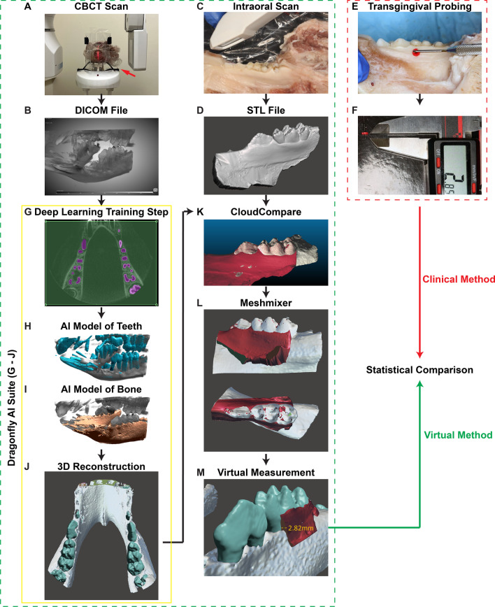
Background: Gingival phenotype plays an important role in dental diagnosis and treatment planning. Traditionally, determining the gingival phenotype is done by manual probing of the gingival soft tissues, an invasive and time-consuming procedure. This study aims to evaluate the feasibility and accuracy of an alternatively novel, non-invasive technology based on the precise 3-dimension (3D) soft tissue reconstruction from intraoral scanning and cone beam computed tomography (CBCT) to predict the gingival biotype.
Methods: As a proof-of-concept, Yorkshire pig mandibles were scanned, and the CBCT data were fed into a deep-learning model to reconstruct the teeth and surrounding bone structure in 3D. By overlaying the CBCT scan with the intraoral scans, an accurate superposition was created and used for virtual measurements of the soft tissue thickness. Meanwhile, gingival thicknesses were also measured by a periodontal probe and digital caliper on the buccal and lingual sides at 3 mm apical to the gingival margin of the posterior teeth and compared with the virtual assessment at the same location. The data obtained from virtual and clinical measurements were compared by Wilcoxon matched-pairs signed-rank analysis, while their correlation was determined by Pearson's r value. The Mann-Whitney U test was used for intergroup comparisons of the amount of difference.
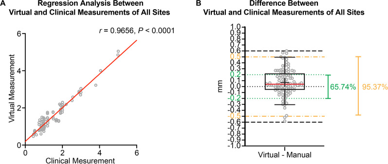
Results: Among 108 investigated locations, the clinical and virtual measurements are strongly positively correlated (r = 0.9656, P < 0.0001), and only clinically insignificant differences (0.066 ± 0.223 mm) were observed between the two assessments. There is no difference in the agreement between the virtual and clinical measurements on sexually matured samples (0.087 ± 0.240 mm) and pre-pubertal samples (0.033 ± 0.195 mm). Noticeably, there is a greater agreement between the virtual and clinical measurements at the buccal sites (0.019 ± 0.233 mm) than at the lingual sites (0.116 ± 0.215 mm).
Conclusion: In summary, the artificial intelligence-based virtual measurement proposed in this work provides an innovative technique potentially for accurately measuring soft tissue thickness using clinical routine 3D imaging systems, which will aid clinicians in generating a more comprehensive diagnosis with less invasive procedures and, in turn, optimize the treatment plans with more predictable outcomes.
期刊介绍:
Progress in Orthodontics is a fully open access, international journal owned by the Italian Society of Orthodontics and published under the brand SpringerOpen. The Society is currently covering all publication costs so there are no article processing charges for authors.
It is a premier journal of international scope that fosters orthodontic research, including both basic research and development of innovative clinical techniques, with an emphasis on the following areas:
• Mechanisms to improve orthodontics
• Clinical studies and control animal studies
• Orthodontics and genetics, genomics
• Temporomandibular joint (TMJ) control clinical trials
• Efficacy of orthodontic appliances and animal models
• Systematic reviews and meta analyses
• Mechanisms to speed orthodontic treatment
Progress in Orthodontics will consider for publication only meritorious and original contributions. These may be:
• Original articles reporting the findings of clinical trials, clinically relevant basic scientific investigations, or novel therapeutic or diagnostic systems
• Review articles on current topics
• Articles on novel techniques and clinical tools
• Articles of contemporary interest


 分享
分享
 求助内容:
求助内容: 应助结果提醒方式:
 扫码关注我们
