{"title":"飞秒激光辅助晶状体角膜内角膜移植术(LIKE)后角膜后表面的变化,将其置入口袋(SMI-LIKE)或皮瓣下(FS-LIKE)。","authors":"Shengtao Liu, Lanhui Yu, Yu Zhao, Xingtao Zhou","doi":"10.1186/s40662-023-00337-2","DOIUrl":null,"url":null,"abstract":"<p><strong>Background: </strong>To compare the changes in posterior corneal surface after small-incision lenticule intrastromal keratoplasty (SMI-LIKE) and femtosecond laser-assisted lenticule intrastromal keratoplasty (FS-LIKE) for hyperopia correction.</p><p><strong>Methods: </strong>In this prospective comparative randomized study, 23 eyes with hyperopia were recruited. Eyes were categorized into two groups-SMI-LIKE group (11 eyes) and FS-LIKE group (12 eyes). Lenticules from myopia small incision lenticule extraction were implanted into a pocket (SMI-LIKE group) or at a depth of 100 µm under a flap (FS-LIKE group). Posterior corneal elevations in the center, mid-periphery, and periphery, as well as mean keratometry of the posterior corneal surface (Kmb) were measured using a Pentacam over a three-month follow-up.</p><p><strong>Results: </strong>All surgeries were completed successfully and no complications occurred. At one day postoperatively, there was a slight backward change with SMI-LIKE and a forward change with FS-LIKE in the central region of the posterior corneal elevation. Conversely, the peripheral area showed forward displacement in SMI-LIKE and an apparent backward change in FS-LIKE. The mid-peripheral regions manifested a backward change after the procedure throughout the entire follow-up in both groups. Kmb exhibited flattening at one month postoperatively and subsequently returned to its original level at three months after SMI-LIKE while in FS-LIKE, Kmb steepened after lenticule implantation with a significant change noted at one day postoperatively (P = 0.001).</p><p><strong>Conclusions: </strong>Posterior corneal surface after SMI-LIKE and FS-LIKE exhibited different change patterns in various corneal regions, with the most prominent change occurring at one day postoperatively during the three-month follow-up.</p><p><strong>Trial registration: </strong>Chinese Clinical Trial Registry: ChiCTR-ONC-16008300. Registered on Apr 18th, 2016. http://www.chictr.org.cn/edit.aspx?pid=14090&htm=4.</p>","PeriodicalId":73010,"journal":{"name":"","volume":"10 1","pages":"23"},"PeriodicalIF":0.0,"publicationDate":"2023-05-01","publicationTypes":"Journal Article","fieldsOfStudy":null,"isOpenAccess":false,"openAccessPdf":"https://www.ncbi.nlm.nih.gov/pmc/articles/PMC10150533/pdf/","citationCount":"0","resultStr":"{\"title\":\"Changes in the posterior corneal surface after femtosecond laser-assisted lenticule intrastromal keratoplasty (LIKE) performed into a pocket (SMI-LIKE) or under a flap (FS-LIKE).\",\"authors\":\"Shengtao Liu, Lanhui Yu, Yu Zhao, Xingtao Zhou\",\"doi\":\"10.1186/s40662-023-00337-2\",\"DOIUrl\":null,\"url\":null,\"abstract\":\"<p><strong>Background: </strong>To compare the changes in posterior corneal surface after small-incision lenticule intrastromal keratoplasty (SMI-LIKE) and femtosecond laser-assisted lenticule intrastromal keratoplasty (FS-LIKE) for hyperopia correction.</p><p><strong>Methods: </strong>In this prospective comparative randomized study, 23 eyes with hyperopia were recruited. Eyes were categorized into two groups-SMI-LIKE group (11 eyes) and FS-LIKE group (12 eyes). Lenticules from myopia small incision lenticule extraction were implanted into a pocket (SMI-LIKE group) or at a depth of 100 µm under a flap (FS-LIKE group). Posterior corneal elevations in the center, mid-periphery, and periphery, as well as mean keratometry of the posterior corneal surface (Kmb) were measured using a Pentacam over a three-month follow-up.</p><p><strong>Results: </strong>All surgeries were completed successfully and no complications occurred. At one day postoperatively, there was a slight backward change with SMI-LIKE and a forward change with FS-LIKE in the central region of the posterior corneal elevation. Conversely, the peripheral area showed forward displacement in SMI-LIKE and an apparent backward change in FS-LIKE. The mid-peripheral regions manifested a backward change after the procedure throughout the entire follow-up in both groups. Kmb exhibited flattening at one month postoperatively and subsequently returned to its original level at three months after SMI-LIKE while in FS-LIKE, Kmb steepened after lenticule implantation with a significant change noted at one day postoperatively (P = 0.001).</p><p><strong>Conclusions: </strong>Posterior corneal surface after SMI-LIKE and FS-LIKE exhibited different change patterns in various corneal regions, with the most prominent change occurring at one day postoperatively during the three-month follow-up.</p><p><strong>Trial registration: </strong>Chinese Clinical Trial Registry: ChiCTR-ONC-16008300. Registered on Apr 18th, 2016. http://www.chictr.org.cn/edit.aspx?pid=14090&htm=4.</p>\",\"PeriodicalId\":73010,\"journal\":{\"name\":\"\",\"volume\":\"10 1\",\"pages\":\"23\"},\"PeriodicalIF\":0.0,\"publicationDate\":\"2023-05-01\",\"publicationTypes\":\"Journal Article\",\"fieldsOfStudy\":null,\"isOpenAccess\":false,\"openAccessPdf\":\"https://www.ncbi.nlm.nih.gov/pmc/articles/PMC10150533/pdf/\",\"citationCount\":\"0\",\"resultStr\":null,\"platform\":\"Semanticscholar\",\"paperid\":null,\"PeriodicalName\":\"\",\"FirstCategoryId\":\"3\",\"ListUrlMain\":\"https://doi.org/10.1186/s40662-023-00337-2\",\"RegionNum\":0,\"RegionCategory\":null,\"ArticlePicture\":[],\"TitleCN\":null,\"AbstractTextCN\":null,\"PMCID\":null,\"EPubDate\":\"\",\"PubModel\":\"\",\"JCR\":\"\",\"JCRName\":\"\",\"Score\":null,\"Total\":0}","platform":"Semanticscholar","paperid":null,"PeriodicalName":"","FirstCategoryId":"3","ListUrlMain":"https://doi.org/10.1186/s40662-023-00337-2","RegionNum":0,"RegionCategory":null,"ArticlePicture":[],"TitleCN":null,"AbstractTextCN":null,"PMCID":null,"EPubDate":"","PubModel":"","JCR":"","JCRName":"","Score":null,"Total":0}
Changes in the posterior corneal surface after femtosecond laser-assisted lenticule intrastromal keratoplasty (LIKE) performed into a pocket (SMI-LIKE) or under a flap (FS-LIKE).
Background: To compare the changes in posterior corneal surface after small-incision lenticule intrastromal keratoplasty (SMI-LIKE) and femtosecond laser-assisted lenticule intrastromal keratoplasty (FS-LIKE) for hyperopia correction.
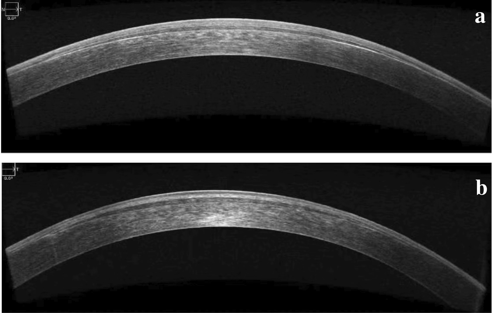
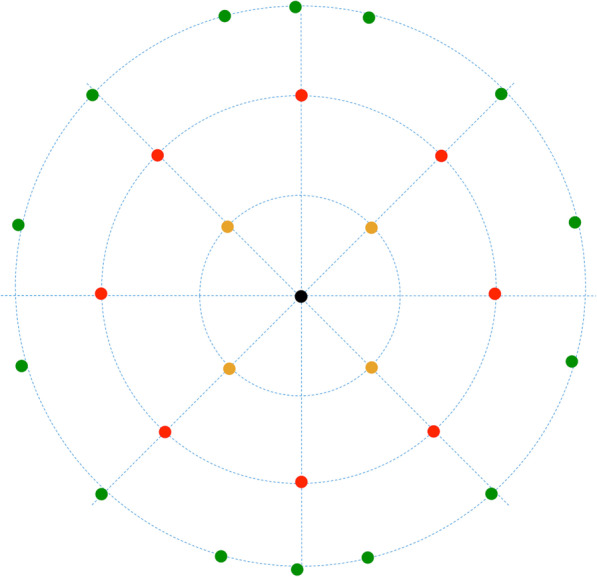
Methods: In this prospective comparative randomized study, 23 eyes with hyperopia were recruited. Eyes were categorized into two groups-SMI-LIKE group (11 eyes) and FS-LIKE group (12 eyes). Lenticules from myopia small incision lenticule extraction were implanted into a pocket (SMI-LIKE group) or at a depth of 100 µm under a flap (FS-LIKE group). Posterior corneal elevations in the center, mid-periphery, and periphery, as well as mean keratometry of the posterior corneal surface (Kmb) were measured using a Pentacam over a three-month follow-up.
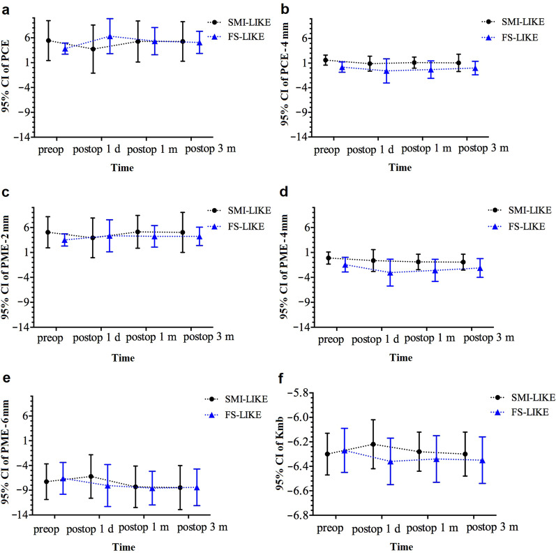
Results: All surgeries were completed successfully and no complications occurred. At one day postoperatively, there was a slight backward change with SMI-LIKE and a forward change with FS-LIKE in the central region of the posterior corneal elevation. Conversely, the peripheral area showed forward displacement in SMI-LIKE and an apparent backward change in FS-LIKE. The mid-peripheral regions manifested a backward change after the procedure throughout the entire follow-up in both groups. Kmb exhibited flattening at one month postoperatively and subsequently returned to its original level at three months after SMI-LIKE while in FS-LIKE, Kmb steepened after lenticule implantation with a significant change noted at one day postoperatively (P = 0.001).
Conclusions: Posterior corneal surface after SMI-LIKE and FS-LIKE exhibited different change patterns in various corneal regions, with the most prominent change occurring at one day postoperatively during the three-month follow-up.
Trial registration: Chinese Clinical Trial Registry: ChiCTR-ONC-16008300. Registered on Apr 18th, 2016. http://www.chictr.org.cn/edit.aspx?pid=14090&htm=4.


 分享
分享
 求助内容:
求助内容: 应助结果提醒方式:
 扫码关注我们
