Kishan Patel, Seung Min Son, Qiwen Zhang, Jeffrey C Wang, Zorica Buser
{"title":"关于直立磁共振成像中沉积征与腰椎间盘突出症之间关系的研究","authors":"Kishan Patel, Seung Min Son, Qiwen Zhang, Jeffrey C Wang, Zorica Buser","doi":"10.1177/21925682231170612","DOIUrl":null,"url":null,"abstract":"<p><strong>Study design: </strong>Retrospective Upright MRI Study.</p><p><strong>Objectives: </strong>Determine the relationship between lumbar disc herniation and presence of the nerve root sedimentation sign on upright kinematic MRI patients.</p><p><strong>Methods: </strong>T2-weighted axial upright kMRI images of 100 patients with the presence of disc herniation in at least 1 lumbar disc between L1/L2 and L5/S1 were obtained. Sedimentation sign, spinal canal anterior-posterior (AP) diameter, disc height, disc herniation size, type of herniation, and zone of herniation were evaluated. A positive sedimentation sign was defined as having either the majority of nerve roots running ventrally or centrally in the canal or conglomeration of the nerve roots at the mid-disc level. Herniation types were defined as either no herniation, disc bulge, protrusion, extrusion, or sequestration. Zones of herniation were categorized as either central, lateral, or far lateral.</p><p><strong>Results: </strong>The kappa value of intra-observer reliability was .915. The kappa value of disc levels with a negative sedimentation sign were seen more frequently (n = 326, 65.2%) than those with a positive sedimentation sign (n = 174, 34.8%). The spinal canal AP diameter was significantly decreased at the L3/L4 and L4/L5 level in patients with a positive sedimentation sign. Discs with a positive sedimentation sign had a larger average size of disc herniation compared to those with a negative sign at all levels. A relationship between positivity of the sedimentation sign and disc herniation type was significant at L2/L3, L3/L4, and L4/L5.</p><p><strong>Conclusions: </strong>Patients with a positive sedimentation sign were seen to have larger disc herniations and more severely degenerated discs.</p>","PeriodicalId":12680,"journal":{"name":"Global Spine Journal","volume":" ","pages":"2088-2094"},"PeriodicalIF":3.0000,"publicationDate":"2024-09-01","publicationTypes":"Journal Article","fieldsOfStudy":null,"isOpenAccess":false,"openAccessPdf":"https://www.ncbi.nlm.nih.gov/pmc/articles/PMC11418682/pdf/","citationCount":"0","resultStr":"{\"title\":\"An Investigation Into the Relationship Between the Sedimentation Sign and Lumbar Disc Herniation in Upright Magnetic Resonance Images.\",\"authors\":\"Kishan Patel, Seung Min Son, Qiwen Zhang, Jeffrey C Wang, Zorica Buser\",\"doi\":\"10.1177/21925682231170612\",\"DOIUrl\":null,\"url\":null,\"abstract\":\"<p><strong>Study design: </strong>Retrospective Upright MRI Study.</p><p><strong>Objectives: </strong>Determine the relationship between lumbar disc herniation and presence of the nerve root sedimentation sign on upright kinematic MRI patients.</p><p><strong>Methods: </strong>T2-weighted axial upright kMRI images of 100 patients with the presence of disc herniation in at least 1 lumbar disc between L1/L2 and L5/S1 were obtained. Sedimentation sign, spinal canal anterior-posterior (AP) diameter, disc height, disc herniation size, type of herniation, and zone of herniation were evaluated. A positive sedimentation sign was defined as having either the majority of nerve roots running ventrally or centrally in the canal or conglomeration of the nerve roots at the mid-disc level. Herniation types were defined as either no herniation, disc bulge, protrusion, extrusion, or sequestration. Zones of herniation were categorized as either central, lateral, or far lateral.</p><p><strong>Results: </strong>The kappa value of intra-observer reliability was .915. The kappa value of disc levels with a negative sedimentation sign were seen more frequently (n = 326, 65.2%) than those with a positive sedimentation sign (n = 174, 34.8%). The spinal canal AP diameter was significantly decreased at the L3/L4 and L4/L5 level in patients with a positive sedimentation sign. Discs with a positive sedimentation sign had a larger average size of disc herniation compared to those with a negative sign at all levels. A relationship between positivity of the sedimentation sign and disc herniation type was significant at L2/L3, L3/L4, and L4/L5.</p><p><strong>Conclusions: </strong>Patients with a positive sedimentation sign were seen to have larger disc herniations and more severely degenerated discs.</p>\",\"PeriodicalId\":12680,\"journal\":{\"name\":\"Global Spine Journal\",\"volume\":\" \",\"pages\":\"2088-2094\"},\"PeriodicalIF\":3.0000,\"publicationDate\":\"2024-09-01\",\"publicationTypes\":\"Journal Article\",\"fieldsOfStudy\":null,\"isOpenAccess\":false,\"openAccessPdf\":\"https://www.ncbi.nlm.nih.gov/pmc/articles/PMC11418682/pdf/\",\"citationCount\":\"0\",\"resultStr\":null,\"platform\":\"Semanticscholar\",\"paperid\":null,\"PeriodicalName\":\"Global Spine Journal\",\"FirstCategoryId\":\"3\",\"ListUrlMain\":\"https://doi.org/10.1177/21925682231170612\",\"RegionNum\":3,\"RegionCategory\":\"医学\",\"ArticlePicture\":[],\"TitleCN\":null,\"AbstractTextCN\":null,\"PMCID\":null,\"EPubDate\":\"2023/4/20 0:00:00\",\"PubModel\":\"Epub\",\"JCR\":\"Q2\",\"JCRName\":\"CLINICAL NEUROLOGY\",\"Score\":null,\"Total\":0}","platform":"Semanticscholar","paperid":null,"PeriodicalName":"Global Spine Journal","FirstCategoryId":"3","ListUrlMain":"https://doi.org/10.1177/21925682231170612","RegionNum":3,"RegionCategory":"医学","ArticlePicture":[],"TitleCN":null,"AbstractTextCN":null,"PMCID":null,"EPubDate":"2023/4/20 0:00:00","PubModel":"Epub","JCR":"Q2","JCRName":"CLINICAL NEUROLOGY","Score":null,"Total":0}
An Investigation Into the Relationship Between the Sedimentation Sign and Lumbar Disc Herniation in Upright Magnetic Resonance Images.
Study design: Retrospective Upright MRI Study.
Objectives: Determine the relationship between lumbar disc herniation and presence of the nerve root sedimentation sign on upright kinematic MRI patients.
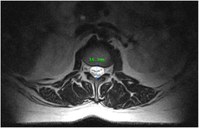
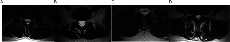
Methods: T2-weighted axial upright kMRI images of 100 patients with the presence of disc herniation in at least 1 lumbar disc between L1/L2 and L5/S1 were obtained. Sedimentation sign, spinal canal anterior-posterior (AP) diameter, disc height, disc herniation size, type of herniation, and zone of herniation were evaluated. A positive sedimentation sign was defined as having either the majority of nerve roots running ventrally or centrally in the canal or conglomeration of the nerve roots at the mid-disc level. Herniation types were defined as either no herniation, disc bulge, protrusion, extrusion, or sequestration. Zones of herniation were categorized as either central, lateral, or far lateral.
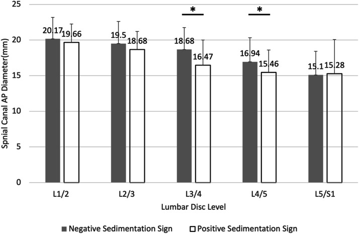
Results: The kappa value of intra-observer reliability was .915. The kappa value of disc levels with a negative sedimentation sign were seen more frequently (n = 326, 65.2%) than those with a positive sedimentation sign (n = 174, 34.8%). The spinal canal AP diameter was significantly decreased at the L3/L4 and L4/L5 level in patients with a positive sedimentation sign. Discs with a positive sedimentation sign had a larger average size of disc herniation compared to those with a negative sign at all levels. A relationship between positivity of the sedimentation sign and disc herniation type was significant at L2/L3, L3/L4, and L4/L5.
Conclusions: Patients with a positive sedimentation sign were seen to have larger disc herniations and more severely degenerated discs.
期刊介绍:
Global Spine Journal (GSJ) is the official scientific publication of AOSpine. A peer-reviewed, open access journal, devoted to the study and treatment of spinal disorders, including diagnosis, operative and non-operative treatment options, surgical techniques, and emerging research and clinical developments.GSJ is indexed in PubMedCentral, SCOPUS, and Emerging Sources Citation Index (ESCI).


 分享
分享
 求助内容:
求助内容: 应助结果提醒方式:
 扫码关注我们
