Sangkyung Choen, Michael S Kent, Abhijit J Chaudhari, Simon R Cherry, Ana Krtolica, Allison L Zwingenberger
{"title":"缺氧示踪剂[18F]FMISO和[18F]FAZA在犬自发性肿瘤中的动态PET/CT成像动力学评价","authors":"Sangkyung Choen, Michael S Kent, Abhijit J Chaudhari, Simon R Cherry, Ana Krtolica, Allison L Zwingenberger","doi":"10.1007/s13139-022-00780-4","DOIUrl":null,"url":null,"abstract":"<p><strong>Purpose: </strong>We evaluated the kinetics of the hypoxia PET radiotracers, [18F]fluoromisonidazole ([18F]FMISO) and [18F]fluoroazomycin-arabinoside ([18F]FAZA), for tumor hypoxia detection and to assess the correlation of hypoxic kinetic parameters with static imaging measures in canine spontaneous tumors.</p><p><strong>Methods: </strong>Sixteen dogs with spontaneous tumors underwent a 150-min dynamic PET scan using either [18F]FMISO or [18F]FAZA. The maximum tumor-to-muscle ratio (TMR<sub>max</sub>) > 1.4 on the last image frame was used as the standard threshold to determine tumor hypoxia. The tumor time-activity curves were analyzed using irreversible and reversible two-tissue compartment models and graphical methods. TMR<sub>max</sub> was compared with radiotracer trapping rate (<i>k</i> <sub>3</sub>), influx rate (<i>K</i> <sub>i</sub>), and distribution volume (<i>V</i> <sub>T</sub>).</p><p><strong>Results: </strong>Tumor hypoxia was detected in 7/8 tumors in the [18F]FMISO group and 4/8 tumors in the [18F]FAZA group. All hypoxic tumors were detected at > 120 min with [18F]FMISO and at > 60 min with [18F]FAZA. [18F]FAZA showed better fit with the reversible model. TMR<sub>max</sub> was strongly correlated with the irreversible parameters (<i>k</i> <sub>3</sub> and <i>K</i> <sub>i</sub>) for [18F]FMISO at > 90 min and with the reversible parameter (<i>V</i> <sub>T</sub>) for [18F]FAZA at > 120 min.</p><p><strong>Conclusions: </strong>Our results showed that [18F]FAZA provided a promising alternative radiotracer to [18F]FMISO with detecting the presence of tumor hypoxia at an earlier time (60 min), consistent with its favorable faster kinetics. The strong correlation between TMR<sub>max</sub> over the 90-150 min and 120-150 min timeframes with [18F]FMISO and [18F]FAZA, respectively, with kinetic parameters associated with tumor hypoxia for each radiotracer, suggests that a static scan measurement (TMR<sub>max</sub>) is a good alternative to quantify tumor hypoxia.</p><p><strong>Supplementary information: </strong>The online version contains supplementary material available at 10.1007/s13139-022-00780-4.</p>","PeriodicalId":19384,"journal":{"name":"Nuclear Medicine and Molecular Imaging","volume":"57 1","pages":"16-25"},"PeriodicalIF":2.7000,"publicationDate":"2023-02-01","publicationTypes":"Journal Article","fieldsOfStudy":null,"isOpenAccess":false,"openAccessPdf":"https://www.ncbi.nlm.nih.gov/pmc/articles/PMC9832187/pdf/","citationCount":"0","resultStr":"{\"title\":\"Kinetic Evaluation of the Hypoxia Radiotracers [<sup>18</sup>F]FMISO and [<sup>18</sup>F]FAZA in Dogs with Spontaneous Tumors Using Dynamic PET/CT Imaging.\",\"authors\":\"Sangkyung Choen, Michael S Kent, Abhijit J Chaudhari, Simon R Cherry, Ana Krtolica, Allison L Zwingenberger\",\"doi\":\"10.1007/s13139-022-00780-4\",\"DOIUrl\":null,\"url\":null,\"abstract\":\"<p><strong>Purpose: </strong>We evaluated the kinetics of the hypoxia PET radiotracers, [18F]fluoromisonidazole ([18F]FMISO) and [18F]fluoroazomycin-arabinoside ([18F]FAZA), for tumor hypoxia detection and to assess the correlation of hypoxic kinetic parameters with static imaging measures in canine spontaneous tumors.</p><p><strong>Methods: </strong>Sixteen dogs with spontaneous tumors underwent a 150-min dynamic PET scan using either [18F]FMISO or [18F]FAZA. The maximum tumor-to-muscle ratio (TMR<sub>max</sub>) > 1.4 on the last image frame was used as the standard threshold to determine tumor hypoxia. The tumor time-activity curves were analyzed using irreversible and reversible two-tissue compartment models and graphical methods. TMR<sub>max</sub> was compared with radiotracer trapping rate (<i>k</i> <sub>3</sub>), influx rate (<i>K</i> <sub>i</sub>), and distribution volume (<i>V</i> <sub>T</sub>).</p><p><strong>Results: </strong>Tumor hypoxia was detected in 7/8 tumors in the [18F]FMISO group and 4/8 tumors in the [18F]FAZA group. All hypoxic tumors were detected at > 120 min with [18F]FMISO and at > 60 min with [18F]FAZA. [18F]FAZA showed better fit with the reversible model. TMR<sub>max</sub> was strongly correlated with the irreversible parameters (<i>k</i> <sub>3</sub> and <i>K</i> <sub>i</sub>) for [18F]FMISO at > 90 min and with the reversible parameter (<i>V</i> <sub>T</sub>) for [18F]FAZA at > 120 min.</p><p><strong>Conclusions: </strong>Our results showed that [18F]FAZA provided a promising alternative radiotracer to [18F]FMISO with detecting the presence of tumor hypoxia at an earlier time (60 min), consistent with its favorable faster kinetics. The strong correlation between TMR<sub>max</sub> over the 90-150 min and 120-150 min timeframes with [18F]FMISO and [18F]FAZA, respectively, with kinetic parameters associated with tumor hypoxia for each radiotracer, suggests that a static scan measurement (TMR<sub>max</sub>) is a good alternative to quantify tumor hypoxia.</p><p><strong>Supplementary information: </strong>The online version contains supplementary material available at 10.1007/s13139-022-00780-4.</p>\",\"PeriodicalId\":19384,\"journal\":{\"name\":\"Nuclear Medicine and Molecular Imaging\",\"volume\":\"57 1\",\"pages\":\"16-25\"},\"PeriodicalIF\":2.7000,\"publicationDate\":\"2023-02-01\",\"publicationTypes\":\"Journal Article\",\"fieldsOfStudy\":null,\"isOpenAccess\":false,\"openAccessPdf\":\"https://www.ncbi.nlm.nih.gov/pmc/articles/PMC9832187/pdf/\",\"citationCount\":\"0\",\"resultStr\":null,\"platform\":\"Semanticscholar\",\"paperid\":null,\"PeriodicalName\":\"Nuclear Medicine and Molecular Imaging\",\"FirstCategoryId\":\"1085\",\"ListUrlMain\":\"https://doi.org/10.1007/s13139-022-00780-4\",\"RegionNum\":0,\"RegionCategory\":null,\"ArticlePicture\":[],\"TitleCN\":null,\"AbstractTextCN\":null,\"PMCID\":null,\"EPubDate\":\"\",\"PubModel\":\"\",\"JCR\":\"Q3\",\"JCRName\":\"RADIOLOGY, NUCLEAR MEDICINE & MEDICAL IMAGING\",\"Score\":null,\"Total\":0}","platform":"Semanticscholar","paperid":null,"PeriodicalName":"Nuclear Medicine and Molecular Imaging","FirstCategoryId":"1085","ListUrlMain":"https://doi.org/10.1007/s13139-022-00780-4","RegionNum":0,"RegionCategory":null,"ArticlePicture":[],"TitleCN":null,"AbstractTextCN":null,"PMCID":null,"EPubDate":"","PubModel":"","JCR":"Q3","JCRName":"RADIOLOGY, NUCLEAR MEDICINE & MEDICAL IMAGING","Score":null,"Total":0}
Kinetic Evaluation of the Hypoxia Radiotracers [18F]FMISO and [18F]FAZA in Dogs with Spontaneous Tumors Using Dynamic PET/CT Imaging.
Purpose: We evaluated the kinetics of the hypoxia PET radiotracers, [18F]fluoromisonidazole ([18F]FMISO) and [18F]fluoroazomycin-arabinoside ([18F]FAZA), for tumor hypoxia detection and to assess the correlation of hypoxic kinetic parameters with static imaging measures in canine spontaneous tumors.
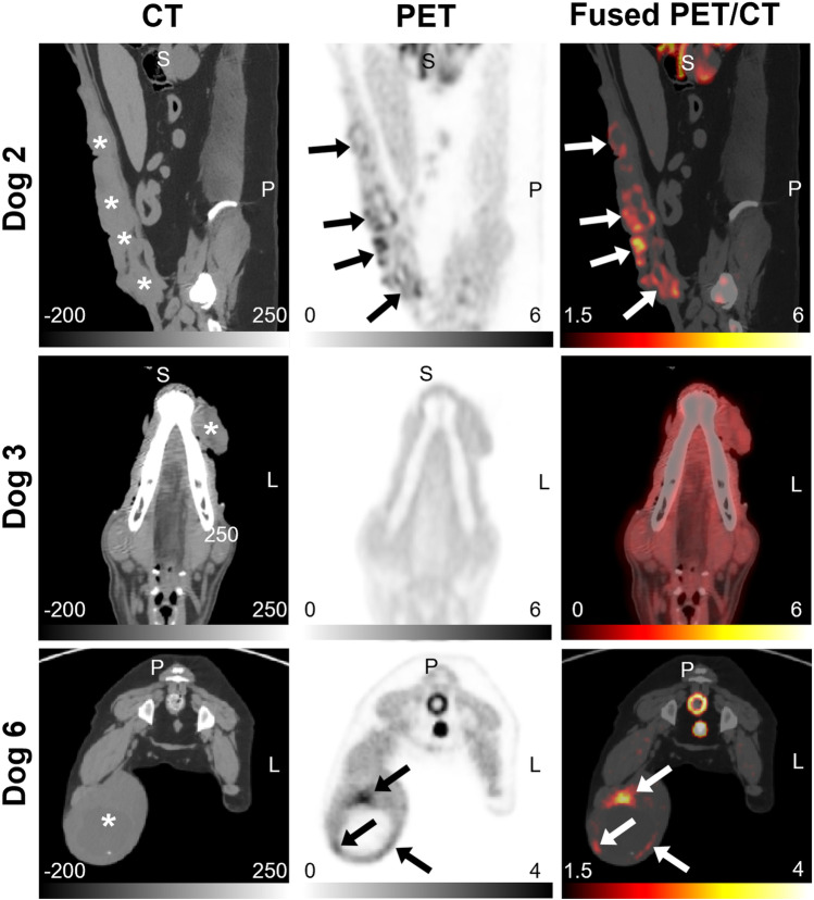
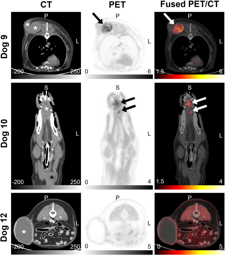
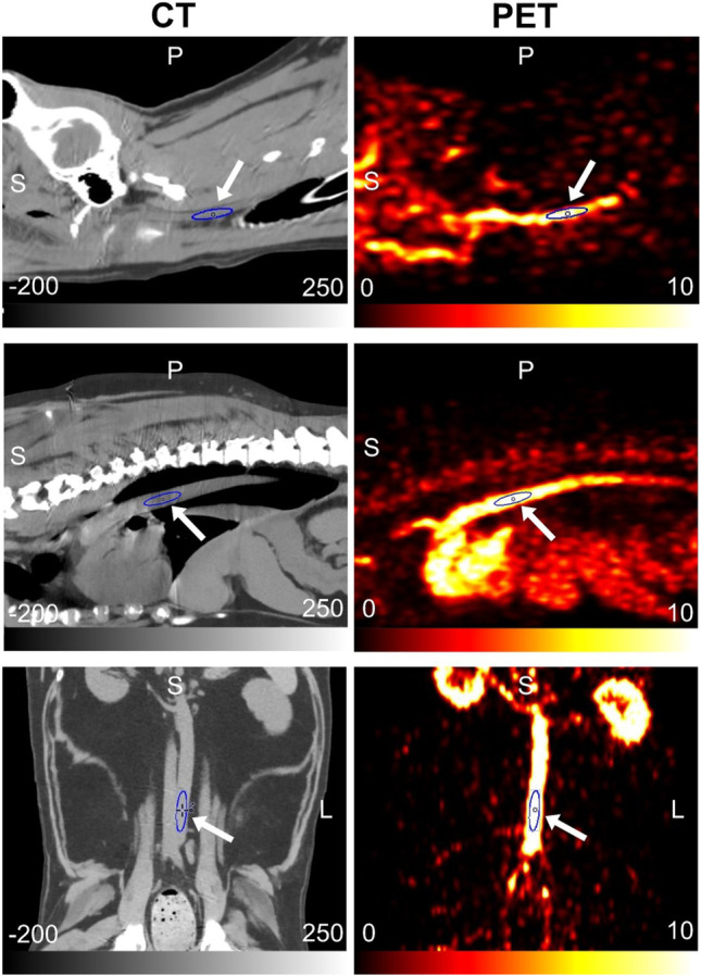
Methods: Sixteen dogs with spontaneous tumors underwent a 150-min dynamic PET scan using either [18F]FMISO or [18F]FAZA. The maximum tumor-to-muscle ratio (TMRmax) > 1.4 on the last image frame was used as the standard threshold to determine tumor hypoxia. The tumor time-activity curves were analyzed using irreversible and reversible two-tissue compartment models and graphical methods. TMRmax was compared with radiotracer trapping rate (k3), influx rate (Ki), and distribution volume (VT).
Results: Tumor hypoxia was detected in 7/8 tumors in the [18F]FMISO group and 4/8 tumors in the [18F]FAZA group. All hypoxic tumors were detected at > 120 min with [18F]FMISO and at > 60 min with [18F]FAZA. [18F]FAZA showed better fit with the reversible model. TMRmax was strongly correlated with the irreversible parameters (k3 and Ki) for [18F]FMISO at > 90 min and with the reversible parameter (VT) for [18F]FAZA at > 120 min.
Conclusions: Our results showed that [18F]FAZA provided a promising alternative radiotracer to [18F]FMISO with detecting the presence of tumor hypoxia at an earlier time (60 min), consistent with its favorable faster kinetics. The strong correlation between TMRmax over the 90-150 min and 120-150 min timeframes with [18F]FMISO and [18F]FAZA, respectively, with kinetic parameters associated with tumor hypoxia for each radiotracer, suggests that a static scan measurement (TMRmax) is a good alternative to quantify tumor hypoxia.
Supplementary information: The online version contains supplementary material available at 10.1007/s13139-022-00780-4.
期刊介绍:
Nuclear Medicine and Molecular Imaging (Nucl Med Mol Imaging) is an official journal of the Korean Society of Nuclear Medicine, which bimonthly publishes papers on February, April, June, August, October, and December about nuclear medicine and related sciences such as radiochemistry, radiopharmacy, dosimetry and pharmacokinetics / pharmacodynamics of radiopharmaceuticals, nuclear and molecular imaging analysis, nuclear and molecular imaging instrumentation, radiation biology and radionuclide therapy. The journal specially welcomes works of artificial intelligence applied to nuclear medicine. The journal will also welcome original works relating to molecular imaging research such as the development of molecular imaging probes, reporter imaging assays, imaging cell trafficking, imaging endo(exo)genous gene expression, and imaging signal transduction. Nucl Med Mol Imaging publishes the following types of papers: original articles, reviews, case reports, editorials, interesting images, and letters to the editor.
The Korean Society of Nuclear Medicine (KSNM)
KSNM is a scientific and professional organization founded in 1961 and a member of the Korean Academy of Medical Sciences of the Korean Medical Association which was established by The Medical Services Law. The aims of KSNM are the promotion of nuclear medicine and cooperation of each member. The business of KSNM includes holding academic meetings and symposia, the publication of journals and books, planning and research of promoting science and health, and training and qualification of nuclear medicine specialists.


 分享
分享
 求助内容:
求助内容: 应助结果提醒方式:
 扫码关注我们
