{"title":"青光眼伴发或并发视网膜静脉闭塞。预测的方面和发展的路径。","authors":"Diana-Maria Dărăbuș, Cristina-Patricia Pac, Mihnea Munteanu","doi":"10.22336/rjo.2023.18","DOIUrl":null,"url":null,"abstract":"<p><p><b>Background and Objectives:</b> The aim of the study is to evaluate prediction factors and progression paths when retinal vein occlusions are associated with preexisting glaucoma or complicated with neovascular glaucoma. <b>Materials and Methods:</b> The study included 111 patients diagnosed with retinal vein occlusions, of whom 21 with preexisting open angle glaucoma and 12 with neovascular glaucoma as complication. The study was conducted from September 2020 to September 2022 in Timişoara, Romania. We assessed intraocular pressure, cup-disc ratio and retinal nerve fiber layer from the moment of retinal vein occlusion diagnosis until at least one year of follow-up, considering these aspects as values of prediction concerning the paths of progression when glaucoma and retinal vein occlusions come together. <b>Results:</b> The mean initial IOP for the affected eyes was higher (15.89 ± 2.73) than for fellow eyes (15.20 ± 3.11), with an increase of the IOP after one year, but with no statistically significant differences for the affected eyes (p=0.116) or for the other eyes (p=0.684), neither for the affected eyes associated with glaucoma in comparison with affected eyes without glaucoma association. The mean cup-disc ratio was higher for the affected eyes in comparison with the fellow eyes (0.4812 ± 0.219 for the affected eyes and 0.4738 ± 0.229 for the fellow ones in cases without associated glaucoma and 0.681 ± 0.157 for the affected eyes and 0.600 ± 0.241 for the fellow eyes in cases with associated glaucoma), with statistical significant differences in the evolution for both groups in comparison with the unaffected eyes (p=0.0056 for the first group and p=0.0003 for the second group). Comparing the evolution of the affected eyes with the preexisting glaucoma and the affected eyes without preexisting glaucoma, no statistical difference has been found (p=0.1104). The mean retinal nerve fiber layer decreased significantly in affected eyes without glaucoma (from 96 ± 14.71 to 89.16 ± 13.07) and in affected eyes with associated glaucoma (from 78.50 ± 4.23 to 75.50 ± 5.83), but with no significant differences (p=0.182). The level of decreasing was significantly more consistent in association with a venous occlusion (p= 0.0001). <b>Conclusions:</b> The findings of the current study fortify the correlation between glaucoma as a risk factor for retinal venous occlusion development, the intraocular pressure and optic nerve cupping as prediction factors in retinal venous occlusions, the association of a well-controlled preexisting glaucoma with no effect on the progression of the retinal venous occlusions and the development of a neovascular glaucoma with a much aggressive and different path of disease progression.</p>","PeriodicalId":21385,"journal":{"name":"Romanian journal of ophthalmology","volume":"67 1","pages":"97-103"},"PeriodicalIF":0.0000,"publicationDate":"2023-01-01","publicationTypes":"Journal Article","fieldsOfStudy":null,"isOpenAccess":false,"openAccessPdf":"https://www.ncbi.nlm.nih.gov/pmc/articles/PMC10117181/pdf/","citationCount":"0","resultStr":"{\"title\":\"Retinal vein occlusions associated or complicated with glaucoma. Aspects of prediction and paths of progression.\",\"authors\":\"Diana-Maria Dărăbuș, Cristina-Patricia Pac, Mihnea Munteanu\",\"doi\":\"10.22336/rjo.2023.18\",\"DOIUrl\":null,\"url\":null,\"abstract\":\"<p><p><b>Background and Objectives:</b> The aim of the study is to evaluate prediction factors and progression paths when retinal vein occlusions are associated with preexisting glaucoma or complicated with neovascular glaucoma. <b>Materials and Methods:</b> The study included 111 patients diagnosed with retinal vein occlusions, of whom 21 with preexisting open angle glaucoma and 12 with neovascular glaucoma as complication. The study was conducted from September 2020 to September 2022 in Timişoara, Romania. We assessed intraocular pressure, cup-disc ratio and retinal nerve fiber layer from the moment of retinal vein occlusion diagnosis until at least one year of follow-up, considering these aspects as values of prediction concerning the paths of progression when glaucoma and retinal vein occlusions come together. <b>Results:</b> The mean initial IOP for the affected eyes was higher (15.89 ± 2.73) than for fellow eyes (15.20 ± 3.11), with an increase of the IOP after one year, but with no statistically significant differences for the affected eyes (p=0.116) or for the other eyes (p=0.684), neither for the affected eyes associated with glaucoma in comparison with affected eyes without glaucoma association. The mean cup-disc ratio was higher for the affected eyes in comparison with the fellow eyes (0.4812 ± 0.219 for the affected eyes and 0.4738 ± 0.229 for the fellow ones in cases without associated glaucoma and 0.681 ± 0.157 for the affected eyes and 0.600 ± 0.241 for the fellow eyes in cases with associated glaucoma), with statistical significant differences in the evolution for both groups in comparison with the unaffected eyes (p=0.0056 for the first group and p=0.0003 for the second group). Comparing the evolution of the affected eyes with the preexisting glaucoma and the affected eyes without preexisting glaucoma, no statistical difference has been found (p=0.1104). The mean retinal nerve fiber layer decreased significantly in affected eyes without glaucoma (from 96 ± 14.71 to 89.16 ± 13.07) and in affected eyes with associated glaucoma (from 78.50 ± 4.23 to 75.50 ± 5.83), but with no significant differences (p=0.182). The level of decreasing was significantly more consistent in association with a venous occlusion (p= 0.0001). <b>Conclusions:</b> The findings of the current study fortify the correlation between glaucoma as a risk factor for retinal venous occlusion development, the intraocular pressure and optic nerve cupping as prediction factors in retinal venous occlusions, the association of a well-controlled preexisting glaucoma with no effect on the progression of the retinal venous occlusions and the development of a neovascular glaucoma with a much aggressive and different path of disease progression.</p>\",\"PeriodicalId\":21385,\"journal\":{\"name\":\"Romanian journal of ophthalmology\",\"volume\":\"67 1\",\"pages\":\"97-103\"},\"PeriodicalIF\":0.0000,\"publicationDate\":\"2023-01-01\",\"publicationTypes\":\"Journal Article\",\"fieldsOfStudy\":null,\"isOpenAccess\":false,\"openAccessPdf\":\"https://www.ncbi.nlm.nih.gov/pmc/articles/PMC10117181/pdf/\",\"citationCount\":\"0\",\"resultStr\":null,\"platform\":\"Semanticscholar\",\"paperid\":null,\"PeriodicalName\":\"Romanian journal of ophthalmology\",\"FirstCategoryId\":\"1085\",\"ListUrlMain\":\"https://doi.org/10.22336/rjo.2023.18\",\"RegionNum\":0,\"RegionCategory\":null,\"ArticlePicture\":[],\"TitleCN\":null,\"AbstractTextCN\":null,\"PMCID\":null,\"EPubDate\":\"\",\"PubModel\":\"\",\"JCR\":\"\",\"JCRName\":\"\",\"Score\":null,\"Total\":0}","platform":"Semanticscholar","paperid":null,"PeriodicalName":"Romanian journal of ophthalmology","FirstCategoryId":"1085","ListUrlMain":"https://doi.org/10.22336/rjo.2023.18","RegionNum":0,"RegionCategory":null,"ArticlePicture":[],"TitleCN":null,"AbstractTextCN":null,"PMCID":null,"EPubDate":"","PubModel":"","JCR":"","JCRName":"","Score":null,"Total":0}
Retinal vein occlusions associated or complicated with glaucoma. Aspects of prediction and paths of progression.
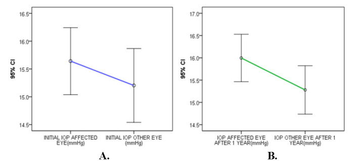
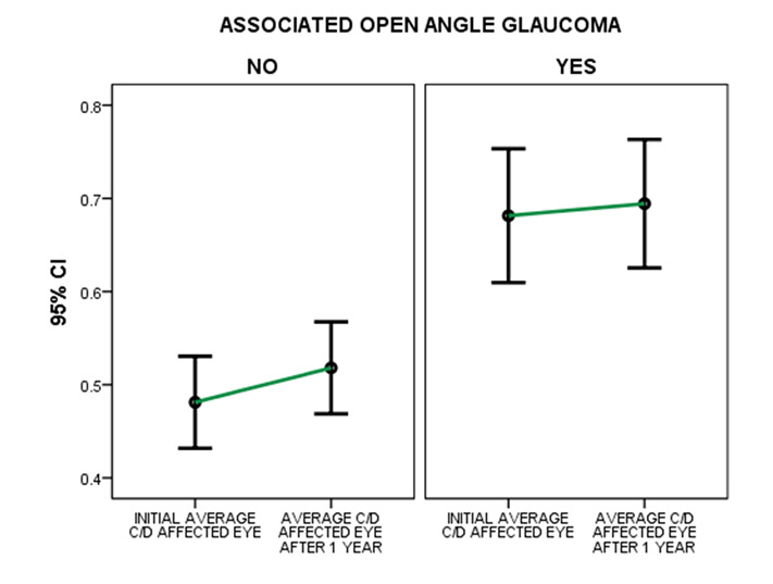
Background and Objectives: The aim of the study is to evaluate prediction factors and progression paths when retinal vein occlusions are associated with preexisting glaucoma or complicated with neovascular glaucoma. Materials and Methods: The study included 111 patients diagnosed with retinal vein occlusions, of whom 21 with preexisting open angle glaucoma and 12 with neovascular glaucoma as complication. The study was conducted from September 2020 to September 2022 in Timişoara, Romania. We assessed intraocular pressure, cup-disc ratio and retinal nerve fiber layer from the moment of retinal vein occlusion diagnosis until at least one year of follow-up, considering these aspects as values of prediction concerning the paths of progression when glaucoma and retinal vein occlusions come together. Results: The mean initial IOP for the affected eyes was higher (15.89 ± 2.73) than for fellow eyes (15.20 ± 3.11), with an increase of the IOP after one year, but with no statistically significant differences for the affected eyes (p=0.116) or for the other eyes (p=0.684), neither for the affected eyes associated with glaucoma in comparison with affected eyes without glaucoma association. The mean cup-disc ratio was higher for the affected eyes in comparison with the fellow eyes (0.4812 ± 0.219 for the affected eyes and 0.4738 ± 0.229 for the fellow ones in cases without associated glaucoma and 0.681 ± 0.157 for the affected eyes and 0.600 ± 0.241 for the fellow eyes in cases with associated glaucoma), with statistical significant differences in the evolution for both groups in comparison with the unaffected eyes (p=0.0056 for the first group and p=0.0003 for the second group). Comparing the evolution of the affected eyes with the preexisting glaucoma and the affected eyes without preexisting glaucoma, no statistical difference has been found (p=0.1104). The mean retinal nerve fiber layer decreased significantly in affected eyes without glaucoma (from 96 ± 14.71 to 89.16 ± 13.07) and in affected eyes with associated glaucoma (from 78.50 ± 4.23 to 75.50 ± 5.83), but with no significant differences (p=0.182). The level of decreasing was significantly more consistent in association with a venous occlusion (p= 0.0001). Conclusions: The findings of the current study fortify the correlation between glaucoma as a risk factor for retinal venous occlusion development, the intraocular pressure and optic nerve cupping as prediction factors in retinal venous occlusions, the association of a well-controlled preexisting glaucoma with no effect on the progression of the retinal venous occlusions and the development of a neovascular glaucoma with a much aggressive and different path of disease progression.


 分享
分享

 求助内容:
求助内容: 应助结果提醒方式:
 扫码关注我们
