{"title":"联合使用细胞阻断和涂片提高细胞学诊断恶性肿瘤的不可触及的乳腺病变的影像学筛查。","authors":"Rieko Nishimura, Mikinao Oiwa","doi":"10.1155/2023/1869858","DOIUrl":null,"url":null,"abstract":"<p><strong>Background: </strong>Currently, core needle biopsy is replacing fine needle aspiration biopsy (FNAB) for pathological diagnosis of breast lesions. However, FNAB is extensively used for diagnosing breast lesions, including screened lesions, at our hospital. Furthermore, direct smears as well as cell blocks (CBs) from the FNAB specimens have been used. To prepare the CBs, hematoxylin and eosin (HE) staining as well as immunostaining with a mixture of p63 and cytokeratin 5/6 antibodies are routinely used. Therefore, in the current study, we sought to assess the efficacy of diagnosing breast lesions using conventional smears and CB immunostaining.</p><p><strong>Methods: </strong>Breast FNAB reports of direct smears and CBs from The Nagoya Medical Center between December 2014 and March 2020, were reviewed. The efficiency of diagnoses made with direct smears and CBs were compared using histology-based diagnoses.</p><p><strong>Results: </strong>Among the 169 histologically confirmed malignant lesions, 12 lesions that were reported as unsatisfactory, benign, or atypia probably benign, using direct smears were diagnosed as malignant using CB. Histologically, these lesions were diagnosed as carcinomas with mild atypia or papillary structures. Ten (83.3%) of the twelve lesions were non-palpable and only detected upon imaging.</p><p><strong>Conclusion: </strong>Combined use of CB and conventional smear leads to the detection of more malignant lesions in breast FNAB specimens, particularly in lesions detected by imaging alone. Immunostaining of CB sections using a mixture of p63 and cytokeratin 5/6 antibodies provides more information than HE staining alone. Breast FNAB with CB preparation can be successfully applied for evaluation of breast lesions in developed countries.</p>","PeriodicalId":49326,"journal":{"name":"Analytical Cellular Pathology","volume":"2023 ","pages":"1869858"},"PeriodicalIF":2.5000,"publicationDate":"2023-01-01","publicationTypes":"Journal Article","fieldsOfStudy":null,"isOpenAccess":false,"openAccessPdf":"https://www.ncbi.nlm.nih.gov/pmc/articles/PMC10171971/pdf/","citationCount":"0","resultStr":"{\"title\":\"Combined Use of Cell Block and Smear Improves the Cytological Diagnosis of Malignancy in Non-Palpable Breast Lesions Screened by Imaging.\",\"authors\":\"Rieko Nishimura, Mikinao Oiwa\",\"doi\":\"10.1155/2023/1869858\",\"DOIUrl\":null,\"url\":null,\"abstract\":\"<p><strong>Background: </strong>Currently, core needle biopsy is replacing fine needle aspiration biopsy (FNAB) for pathological diagnosis of breast lesions. However, FNAB is extensively used for diagnosing breast lesions, including screened lesions, at our hospital. Furthermore, direct smears as well as cell blocks (CBs) from the FNAB specimens have been used. To prepare the CBs, hematoxylin and eosin (HE) staining as well as immunostaining with a mixture of p63 and cytokeratin 5/6 antibodies are routinely used. Therefore, in the current study, we sought to assess the efficacy of diagnosing breast lesions using conventional smears and CB immunostaining.</p><p><strong>Methods: </strong>Breast FNAB reports of direct smears and CBs from The Nagoya Medical Center between December 2014 and March 2020, were reviewed. The efficiency of diagnoses made with direct smears and CBs were compared using histology-based diagnoses.</p><p><strong>Results: </strong>Among the 169 histologically confirmed malignant lesions, 12 lesions that were reported as unsatisfactory, benign, or atypia probably benign, using direct smears were diagnosed as malignant using CB. Histologically, these lesions were diagnosed as carcinomas with mild atypia or papillary structures. Ten (83.3%) of the twelve lesions were non-palpable and only detected upon imaging.</p><p><strong>Conclusion: </strong>Combined use of CB and conventional smear leads to the detection of more malignant lesions in breast FNAB specimens, particularly in lesions detected by imaging alone. Immunostaining of CB sections using a mixture of p63 and cytokeratin 5/6 antibodies provides more information than HE staining alone. Breast FNAB with CB preparation can be successfully applied for evaluation of breast lesions in developed countries.</p>\",\"PeriodicalId\":49326,\"journal\":{\"name\":\"Analytical Cellular Pathology\",\"volume\":\"2023 \",\"pages\":\"1869858\"},\"PeriodicalIF\":2.5000,\"publicationDate\":\"2023-01-01\",\"publicationTypes\":\"Journal Article\",\"fieldsOfStudy\":null,\"isOpenAccess\":false,\"openAccessPdf\":\"https://www.ncbi.nlm.nih.gov/pmc/articles/PMC10171971/pdf/\",\"citationCount\":\"0\",\"resultStr\":null,\"platform\":\"Semanticscholar\",\"paperid\":null,\"PeriodicalName\":\"Analytical Cellular Pathology\",\"FirstCategoryId\":\"3\",\"ListUrlMain\":\"https://doi.org/10.1155/2023/1869858\",\"RegionNum\":4,\"RegionCategory\":\"医学\",\"ArticlePicture\":[],\"TitleCN\":null,\"AbstractTextCN\":null,\"PMCID\":null,\"EPubDate\":\"\",\"PubModel\":\"\",\"JCR\":\"Q3\",\"JCRName\":\"CELL BIOLOGY\",\"Score\":null,\"Total\":0}","platform":"Semanticscholar","paperid":null,"PeriodicalName":"Analytical Cellular Pathology","FirstCategoryId":"3","ListUrlMain":"https://doi.org/10.1155/2023/1869858","RegionNum":4,"RegionCategory":"医学","ArticlePicture":[],"TitleCN":null,"AbstractTextCN":null,"PMCID":null,"EPubDate":"","PubModel":"","JCR":"Q3","JCRName":"CELL BIOLOGY","Score":null,"Total":0}
Combined Use of Cell Block and Smear Improves the Cytological Diagnosis of Malignancy in Non-Palpable Breast Lesions Screened by Imaging.
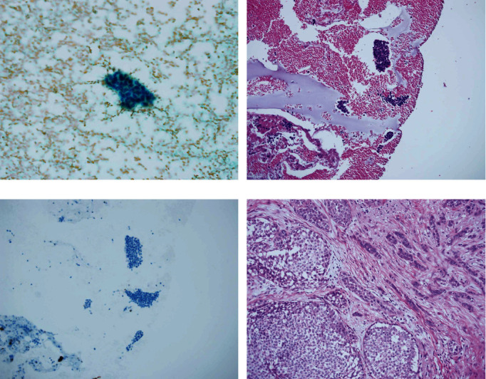
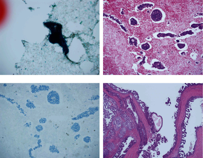
Background: Currently, core needle biopsy is replacing fine needle aspiration biopsy (FNAB) for pathological diagnosis of breast lesions. However, FNAB is extensively used for diagnosing breast lesions, including screened lesions, at our hospital. Furthermore, direct smears as well as cell blocks (CBs) from the FNAB specimens have been used. To prepare the CBs, hematoxylin and eosin (HE) staining as well as immunostaining with a mixture of p63 and cytokeratin 5/6 antibodies are routinely used. Therefore, in the current study, we sought to assess the efficacy of diagnosing breast lesions using conventional smears and CB immunostaining.
Methods: Breast FNAB reports of direct smears and CBs from The Nagoya Medical Center between December 2014 and March 2020, were reviewed. The efficiency of diagnoses made with direct smears and CBs were compared using histology-based diagnoses.
Results: Among the 169 histologically confirmed malignant lesions, 12 lesions that were reported as unsatisfactory, benign, or atypia probably benign, using direct smears were diagnosed as malignant using CB. Histologically, these lesions were diagnosed as carcinomas with mild atypia or papillary structures. Ten (83.3%) of the twelve lesions were non-palpable and only detected upon imaging.
Conclusion: Combined use of CB and conventional smear leads to the detection of more malignant lesions in breast FNAB specimens, particularly in lesions detected by imaging alone. Immunostaining of CB sections using a mixture of p63 and cytokeratin 5/6 antibodies provides more information than HE staining alone. Breast FNAB with CB preparation can be successfully applied for evaluation of breast lesions in developed countries.
期刊介绍:
Analytical Cellular Pathology is a peer-reviewed, Open Access journal that provides a forum for scientists, medical practitioners and pathologists working in the area of cellular pathology. The journal publishes original research articles, review articles, and clinical studies related to cytology, carcinogenesis, cell receptors, biomarkers, diagnostic pathology, immunopathology, and hematology.


 分享
分享
 求助内容:
求助内容: 应助结果提醒方式:
 扫码关注我们
