IF 1.4 3区 医学Q4 BIOCHEMISTRY & MOLECULAR BIOLOGYMolecular VisionPub Date : 2023-05-20eCollection Date: 2023-01-01
Thomas Michael Shiju, Lycia Pedral Sampaio, Guilherme S L Hilgert, Steven E Wilson
{"title":"在伤口愈合过程中,角膜上皮基底膜的组装是由上皮细胞与角膜成纤维细胞协调进行的。","authors":"Thomas Michael Shiju, Lycia Pedral Sampaio, Guilherme S L Hilgert, Steven E Wilson","doi":"","DOIUrl":null,"url":null,"abstract":"<p><strong>Purpose: </strong>To understand which cell types, either alone or in combination, contribute to the assembly of the epithelial basement membrane (BM) during corneal wound healing.</p><p><strong>Methods: </strong>A 3D corneal organotypic model and an in situ rabbit photorefractive keratectomy (PRK) model were used in this study. The 3D corneal organotypic model was established by culturing the rabbit corneal epithelial cells with either corneal fibroblasts or myofibroblasts embedded in collagen type I for 18 days. Corneal fibroblasts were isolated from fresh rabbit corneas, and the myofibroblasts were derived either directly from bone marrow or differentiated from corneal fibroblasts. Immunocytochemistry for alpha-smooth muscle actin (SMA), vimentin, desmin, and vinculin markers confirmed well-differentiated myofibroblasts. Immunohistochemistry was performed in cryofixed sections for BM markers, including laminin alpha-5, laminin beta-3, perlecan, nidogen-1, and collagen type IV. Specimens were also examined with transmission electron microscopy (TEM). Corneas were collected from rabbits after -3 diopter (D) PRK at different time points after surgery, with four corneas at each time point in each group. Cryofixed corneal sections were stained for vimentin, alpha-SMA, and nidogen-1.</p><p><strong>Results: </strong>The formation of an epithelial BM with expression of laminin alpha-5, laminin beta-3, perlecan, nidogen-1, and collagen IV was observed at the interface between the corneal epithelial cells and corneal fibroblasts. TEM images further confirmed the presence of epithelial BM in organotypic cultures of epithelial cells and corneal fibroblasts. No epithelial BM was observed in cultures of corneal epithelial cells and myofibroblasts (cornea or bone marrow derived), corneal epithelial cells alone, or corneal fibroblasts alone. In rabbit corneas after -3D PRK, a strong association was observed between the regenerating epithelial BM and the presence of corneal fibroblasts at the site of epithelial BM generation.</p><p><strong>Conclusions: </strong>The corneal epithelial BM assembly is mediated by epithelial cells in coordination with corneal fibroblasts during wound healing.</p>","PeriodicalId":18866,"journal":{"name":"Molecular Vision","volume":"29 ","pages":"68-86"},"PeriodicalIF":1.4000,"publicationDate":"2023-05-20","publicationTypes":"Journal Article","fieldsOfStudy":null,"isOpenAccess":false,"openAccessPdf":"https://ftp.ncbi.nlm.nih.gov/pub/pmc/oa_pdf/4b/04/mv-v29-68.PMC10243680.pdf","citationCount":"0","resultStr":"{\"title\":\"Corneal epithelial basement membrane assembly is mediated by epithelial cells in coordination with corneal fibroblasts during wound healing.\",\"authors\":\"Thomas Michael Shiju, Lycia Pedral Sampaio, Guilherme S L Hilgert, Steven E Wilson\",\"doi\":\"\",\"DOIUrl\":null,\"url\":null,\"abstract\":\"<p><strong>Purpose: </strong>To understand which cell types, either alone or in combination, contribute to the assembly of the epithelial basement membrane (BM) during corneal wound healing.</p><p><strong>Methods: </strong>A 3D corneal organotypic model and an in situ rabbit photorefractive keratectomy (PRK) model were used in this study. The 3D corneal organotypic model was established by culturing the rabbit corneal epithelial cells with either corneal fibroblasts or myofibroblasts embedded in collagen type I for 18 days. Corneal fibroblasts were isolated from fresh rabbit corneas, and the myofibroblasts were derived either directly from bone marrow or differentiated from corneal fibroblasts. Immunocytochemistry for alpha-smooth muscle actin (SMA), vimentin, desmin, and vinculin markers confirmed well-differentiated myofibroblasts. Immunohistochemistry was performed in cryofixed sections for BM markers, including laminin alpha-5, laminin beta-3, perlecan, nidogen-1, and collagen type IV. Specimens were also examined with transmission electron microscopy (TEM). Corneas were collected from rabbits after -3 diopter (D) PRK at different time points after surgery, with four corneas at each time point in each group. Cryofixed corneal sections were stained for vimentin, alpha-SMA, and nidogen-1.</p><p><strong>Results: </strong>The formation of an epithelial BM with expression of laminin alpha-5, laminin beta-3, perlecan, nidogen-1, and collagen IV was observed at the interface between the corneal epithelial cells and corneal fibroblasts. TEM images further confirmed the presence of epithelial BM in organotypic cultures of epithelial cells and corneal fibroblasts. No epithelial BM was observed in cultures of corneal epithelial cells and myofibroblasts (cornea or bone marrow derived), corneal epithelial cells alone, or corneal fibroblasts alone. In rabbit corneas after -3D PRK, a strong association was observed between the regenerating epithelial BM and the presence of corneal fibroblasts at the site of epithelial BM generation.</p><p><strong>Conclusions: </strong>The corneal epithelial BM assembly is mediated by epithelial cells in coordination with corneal fibroblasts during wound healing.</p>\",\"PeriodicalId\":18866,\"journal\":{\"name\":\"Molecular Vision\",\"volume\":\"29 \",\"pages\":\"68-86\"},\"PeriodicalIF\":1.4000,\"publicationDate\":\"2023-05-20\",\"publicationTypes\":\"Journal Article\",\"fieldsOfStudy\":null,\"isOpenAccess\":false,\"openAccessPdf\":\"https://ftp.ncbi.nlm.nih.gov/pub/pmc/oa_pdf/4b/04/mv-v29-68.PMC10243680.pdf\",\"citationCount\":\"0\",\"resultStr\":null,\"platform\":\"Semanticscholar\",\"paperid\":null,\"PeriodicalName\":\"Molecular Vision\",\"FirstCategoryId\":\"3\",\"ListUrlMain\":\"\",\"RegionNum\":3,\"RegionCategory\":\"医学\",\"ArticlePicture\":[],\"TitleCN\":null,\"AbstractTextCN\":null,\"PMCID\":null,\"EPubDate\":\"2023/1/1 0:00:00\",\"PubModel\":\"eCollection\",\"JCR\":\"Q4\",\"JCRName\":\"BIOCHEMISTRY & MOLECULAR BIOLOGY\",\"Score\":null,\"Total\":0}","platform":"Semanticscholar","paperid":null,"PeriodicalName":"Molecular Vision","FirstCategoryId":"3","ListUrlMain":"","RegionNum":3,"RegionCategory":"医学","ArticlePicture":[],"TitleCN":null,"AbstractTextCN":null,"PMCID":null,"EPubDate":"2023/1/1 0:00:00","PubModel":"eCollection","JCR":"Q4","JCRName":"BIOCHEMISTRY & MOLECULAR BIOLOGY","Score":null,"Total":0}
Corneal epithelial basement membrane assembly is mediated by epithelial cells in coordination with corneal fibroblasts during wound healing.
Purpose: To understand which cell types, either alone or in combination, contribute to the assembly of the epithelial basement membrane (BM) during corneal wound healing.
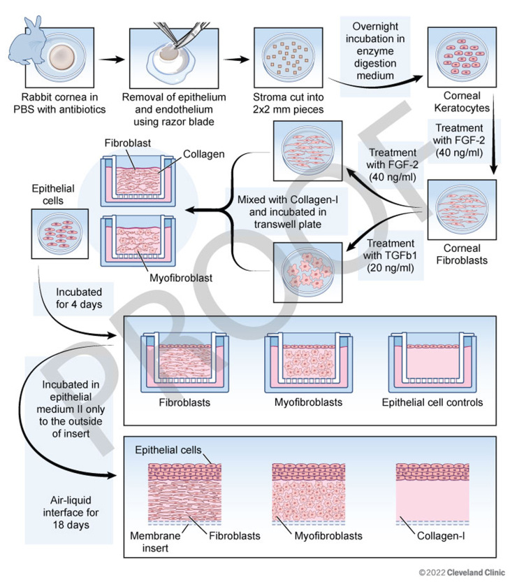
Methods: A 3D corneal organotypic model and an in situ rabbit photorefractive keratectomy (PRK) model were used in this study. The 3D corneal organotypic model was established by culturing the rabbit corneal epithelial cells with either corneal fibroblasts or myofibroblasts embedded in collagen type I for 18 days. Corneal fibroblasts were isolated from fresh rabbit corneas, and the myofibroblasts were derived either directly from bone marrow or differentiated from corneal fibroblasts. Immunocytochemistry for alpha-smooth muscle actin (SMA), vimentin, desmin, and vinculin markers confirmed well-differentiated myofibroblasts. Immunohistochemistry was performed in cryofixed sections for BM markers, including laminin alpha-5, laminin beta-3, perlecan, nidogen-1, and collagen type IV. Specimens were also examined with transmission electron microscopy (TEM). Corneas were collected from rabbits after -3 diopter (D) PRK at different time points after surgery, with four corneas at each time point in each group. Cryofixed corneal sections were stained for vimentin, alpha-SMA, and nidogen-1.
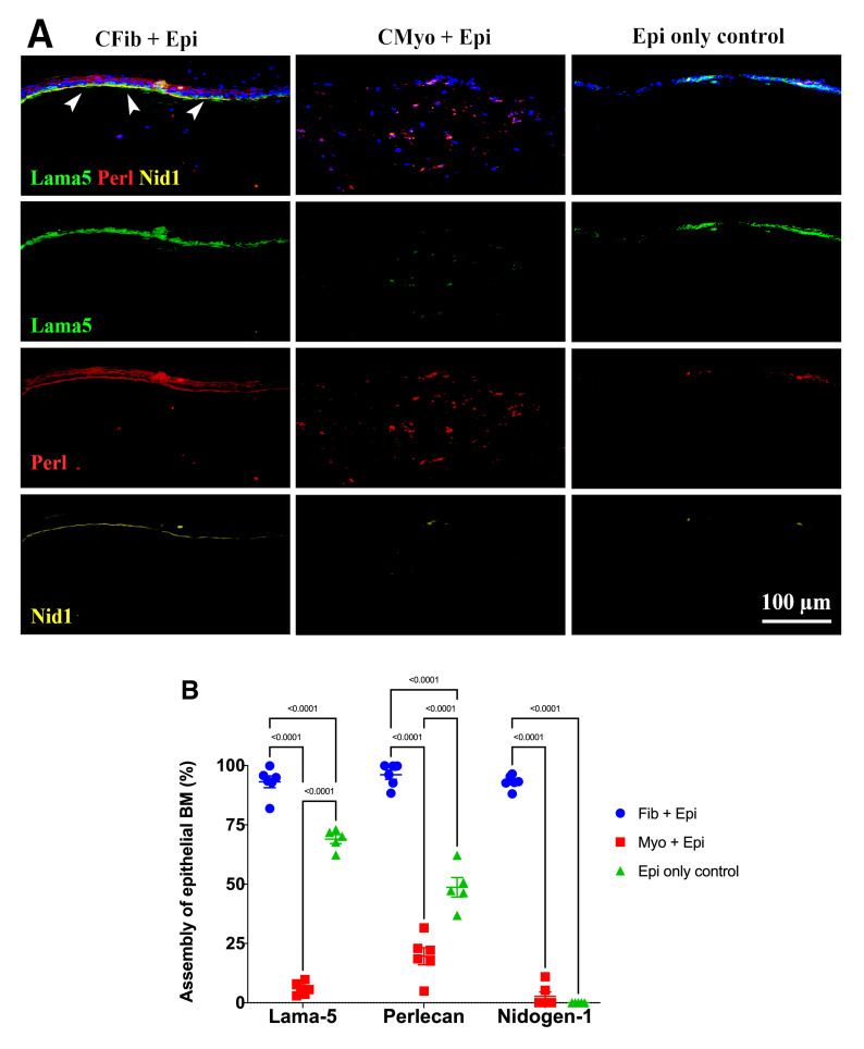
Results: The formation of an epithelial BM with expression of laminin alpha-5, laminin beta-3, perlecan, nidogen-1, and collagen IV was observed at the interface between the corneal epithelial cells and corneal fibroblasts. TEM images further confirmed the presence of epithelial BM in organotypic cultures of epithelial cells and corneal fibroblasts. No epithelial BM was observed in cultures of corneal epithelial cells and myofibroblasts (cornea or bone marrow derived), corneal epithelial cells alone, or corneal fibroblasts alone. In rabbit corneas after -3D PRK, a strong association was observed between the regenerating epithelial BM and the presence of corneal fibroblasts at the site of epithelial BM generation.
Conclusions: The corneal epithelial BM assembly is mediated by epithelial cells in coordination with corneal fibroblasts during wound healing.
期刊介绍:
Molecular Vision is a peer-reviewed journal dedicated to the dissemination of research results in molecular biology, cell biology, and the genetics of the visual system (ocular and cortical).
Molecular Vision publishes articles presenting original research that has not previously been published and comprehensive articles reviewing the current status of a particular field or topic. Submissions to Molecular Vision are subjected to rigorous peer review. Molecular Vision does NOT publish preprints.
For authors, Molecular Vision provides a rapid means of communicating important results. Access to Molecular Vision is free and unrestricted, allowing the widest possible audience for your article. Digital publishing allows you to use color images freely (and without fees). Additionally, you may publish animations, sounds, or other supplementary information that clarifies or supports your article. Each of the authors of an article may also list an electronic mail address (which will be updated upon request) to give interested readers easy access to authors.


 分享
分享


 求助内容:
求助内容: 应助结果提醒方式:
 扫码关注我们
