Theresa T. Cody, Yasunari Kiryu, Micah D. Bakenhaster, Kuttichantran Subramaniam, Maki Tabuchi, Mohammad Shamim Ahasan, Holden E. Harris, Jan H. Landsberg, Thomas B. Waltzek, Alexander Q. Fogg, Colin Shea, Deborah B. Pouder, William F. Patterson III, Meaghan E. Emory, Roy P. Yanong
{"title":"在墨西哥湾影响狮子鱼的皮肤溃疡性病变的未知病因","authors":"Theresa T. Cody, Yasunari Kiryu, Micah D. Bakenhaster, Kuttichantran Subramaniam, Maki Tabuchi, Mohammad Shamim Ahasan, Holden E. Harris, Jan H. Landsberg, Thomas B. Waltzek, Alexander Q. Fogg, Colin Shea, Deborah B. Pouder, William F. Patterson III, Meaghan E. Emory, Roy P. Yanong","doi":"10.1002/aah.10174","DOIUrl":null,"url":null,"abstract":"<div>\n \n \n <section>\n \n <h3> Objective</h3>\n \n <p>Cutaneous ulcerative skin lesions in a complex of invasive Gulf of Mexico lionfish (Red Lionfish <i>Pterois volitans</i>, Devil Firefish <i>P. miles</i>, and the hybrid Red Lionfish × Devil Firefish) became epizootic beginning in mid-August 2017. Herein, we provide the first pathological descriptions of these lesions and summarize our analyses to elucidate the etiology of the disease.</p>\n </section>\n \n <section>\n \n <h3> Methods</h3>\n \n <p>We examined ulcerated and normal fish through gross pathology and histopathology, bacterial sampling, and unbiased metagenomic next-generation sequencing. We tracked prevalence of the disease, and we used biological health indicators (condition factor, splenosomatic and hepatosomatic index) to evaluate impacts to health, while considering sex and age as potential risk factors.</p>\n </section>\n \n <section>\n \n <h3> Result</h3>\n \n <p>Typical ulcerative lesions were deep, exposing skeletal muscle, and were bordered by pale or reddened areas often with some degree of scale loss. Only incidental parasites were found in our examinations. Most fish (86%; <i>n</i> = 50) exhibited wound healing grossly and histologically, confirmed by the presence of granulation tissues. A primary bacterial pathogen was not evident through bacterial culture or histopathology. Metagenomic next-generation sequencing did not reveal a viral pathogen (DNA or RNA) but did provide information about the microbiome of some ulcerated specimens. Compared with clinically healthy fish, ulcerated fish had a significantly lower condition factor and a higher splenosomatic index. Disease prevalence at monitored sites through July 2021 indicated that ulcerated fish were still present but at substantially lower prevalence than observed in 2017.</p>\n </section>\n \n <section>\n \n <h3> Conclusion</h3>\n \n <p>Although some common findings in a number of specimens suggest a potential role for opportunistic bacteria, collectively our suite of diagnostics and analyses did not reveal an intralesional infectious agent, and we must consider the possibility that there was no communicable pathogen.</p>\n </section>\n </div>","PeriodicalId":15235,"journal":{"name":"Journal of aquatic animal health","volume":"35 1","pages":"20-33"},"PeriodicalIF":1.7000,"publicationDate":"2023-01-27","publicationTypes":"Journal Article","fieldsOfStudy":null,"isOpenAccess":false,"openAccessPdf":"https://onlinelibrary.wiley.com/doi/epdf/10.1002/aah.10174","citationCount":"1","resultStr":"{\"title\":\"Cutaneous ulcerative lesions of unknown etiology affecting lionfish Pterois spp. in the Gulf of Mexico\",\"authors\":\"Theresa T. Cody, Yasunari Kiryu, Micah D. Bakenhaster, Kuttichantran Subramaniam, Maki Tabuchi, Mohammad Shamim Ahasan, Holden E. Harris, Jan H. Landsberg, Thomas B. Waltzek, Alexander Q. Fogg, Colin Shea, Deborah B. Pouder, William F. Patterson III, Meaghan E. Emory, Roy P. Yanong\",\"doi\":\"10.1002/aah.10174\",\"DOIUrl\":null,\"url\":null,\"abstract\":\"<div>\\n \\n \\n <section>\\n \\n <h3> Objective</h3>\\n \\n <p>Cutaneous ulcerative skin lesions in a complex of invasive Gulf of Mexico lionfish (Red Lionfish <i>Pterois volitans</i>, Devil Firefish <i>P. miles</i>, and the hybrid Red Lionfish × Devil Firefish) became epizootic beginning in mid-August 2017. Herein, we provide the first pathological descriptions of these lesions and summarize our analyses to elucidate the etiology of the disease.</p>\\n </section>\\n \\n <section>\\n \\n <h3> Methods</h3>\\n \\n <p>We examined ulcerated and normal fish through gross pathology and histopathology, bacterial sampling, and unbiased metagenomic next-generation sequencing. We tracked prevalence of the disease, and we used biological health indicators (condition factor, splenosomatic and hepatosomatic index) to evaluate impacts to health, while considering sex and age as potential risk factors.</p>\\n </section>\\n \\n <section>\\n \\n <h3> Result</h3>\\n \\n <p>Typical ulcerative lesions were deep, exposing skeletal muscle, and were bordered by pale or reddened areas often with some degree of scale loss. Only incidental parasites were found in our examinations. Most fish (86%; <i>n</i> = 50) exhibited wound healing grossly and histologically, confirmed by the presence of granulation tissues. A primary bacterial pathogen was not evident through bacterial culture or histopathology. Metagenomic next-generation sequencing did not reveal a viral pathogen (DNA or RNA) but did provide information about the microbiome of some ulcerated specimens. Compared with clinically healthy fish, ulcerated fish had a significantly lower condition factor and a higher splenosomatic index. Disease prevalence at monitored sites through July 2021 indicated that ulcerated fish were still present but at substantially lower prevalence than observed in 2017.</p>\\n </section>\\n \\n <section>\\n \\n <h3> Conclusion</h3>\\n \\n <p>Although some common findings in a number of specimens suggest a potential role for opportunistic bacteria, collectively our suite of diagnostics and analyses did not reveal an intralesional infectious agent, and we must consider the possibility that there was no communicable pathogen.</p>\\n </section>\\n </div>\",\"PeriodicalId\":15235,\"journal\":{\"name\":\"Journal of aquatic animal health\",\"volume\":\"35 1\",\"pages\":\"20-33\"},\"PeriodicalIF\":1.7000,\"publicationDate\":\"2023-01-27\",\"publicationTypes\":\"Journal Article\",\"fieldsOfStudy\":null,\"isOpenAccess\":false,\"openAccessPdf\":\"https://onlinelibrary.wiley.com/doi/epdf/10.1002/aah.10174\",\"citationCount\":\"1\",\"resultStr\":null,\"platform\":\"Semanticscholar\",\"paperid\":null,\"PeriodicalName\":\"Journal of aquatic animal health\",\"FirstCategoryId\":\"97\",\"ListUrlMain\":\"https://onlinelibrary.wiley.com/doi/10.1002/aah.10174\",\"RegionNum\":4,\"RegionCategory\":\"农林科学\",\"ArticlePicture\":[],\"TitleCN\":null,\"AbstractTextCN\":null,\"PMCID\":null,\"EPubDate\":\"\",\"PubModel\":\"\",\"JCR\":\"Q3\",\"JCRName\":\"FISHERIES\",\"Score\":null,\"Total\":0}","platform":"Semanticscholar","paperid":null,"PeriodicalName":"Journal of aquatic animal health","FirstCategoryId":"97","ListUrlMain":"https://onlinelibrary.wiley.com/doi/10.1002/aah.10174","RegionNum":4,"RegionCategory":"农林科学","ArticlePicture":[],"TitleCN":null,"AbstractTextCN":null,"PMCID":null,"EPubDate":"","PubModel":"","JCR":"Q3","JCRName":"FISHERIES","Score":null,"Total":0}
Cutaneous ulcerative lesions of unknown etiology affecting lionfish Pterois spp. in the Gulf of Mexico
Objective
Cutaneous ulcerative skin lesions in a complex of invasive Gulf of Mexico lionfish (Red Lionfish Pterois volitans, Devil Firefish P. miles, and the hybrid Red Lionfish × Devil Firefish) became epizootic beginning in mid-August 2017. Herein, we provide the first pathological descriptions of these lesions and summarize our analyses to elucidate the etiology of the disease.
Methods
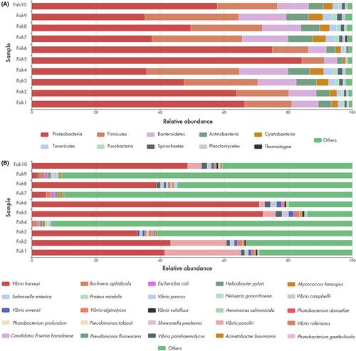
We examined ulcerated and normal fish through gross pathology and histopathology, bacterial sampling, and unbiased metagenomic next-generation sequencing. We tracked prevalence of the disease, and we used biological health indicators (condition factor, splenosomatic and hepatosomatic index) to evaluate impacts to health, while considering sex and age as potential risk factors.
Result
Typical ulcerative lesions were deep, exposing skeletal muscle, and were bordered by pale or reddened areas often with some degree of scale loss. Only incidental parasites were found in our examinations. Most fish (86%; n = 50) exhibited wound healing grossly and histologically, confirmed by the presence of granulation tissues. A primary bacterial pathogen was not evident through bacterial culture or histopathology. Metagenomic next-generation sequencing did not reveal a viral pathogen (DNA or RNA) but did provide information about the microbiome of some ulcerated specimens. Compared with clinically healthy fish, ulcerated fish had a significantly lower condition factor and a higher splenosomatic index. Disease prevalence at monitored sites through July 2021 indicated that ulcerated fish were still present but at substantially lower prevalence than observed in 2017.
Conclusion
Although some common findings in a number of specimens suggest a potential role for opportunistic bacteria, collectively our suite of diagnostics and analyses did not reveal an intralesional infectious agent, and we must consider the possibility that there was no communicable pathogen.
期刊介绍:
The Journal of Aquatic Animal Health serves the international community of scientists and culturists concerned with the health of aquatic organisms. It carries research papers on the causes, effects, treatments, and prevention of diseases of marine and freshwater organisms, particularly fish and shellfish. In addition, it contains papers that describe biochemical and physiological investigations into fish health that relate to assessing the impacts of both environmental and pathogenic features.


 分享
分享
 求助内容:
求助内容: 应助结果提醒方式:
 扫码关注我们
