Patricia J McLaughlin, Joseph W Sassani, David Diaz, Ian S Zagon
{"title":"阿片生长因子升高改变1型糖尿病大鼠的边缘。","authors":"Patricia J McLaughlin, Joseph W Sassani, David Diaz, Ian S Zagon","doi":"10.33696/diabetes.4.054","DOIUrl":null,"url":null,"abstract":"<p><p>Ocular surface complications occur in more than 50% of individuals diagnosed with diabetes. The financial and health-related burden of diabetes is increasing annually. Several major ocular complications associated with diabetes involve the limbus. The vascular limbus, adjacent to the avascular cornea, is the source of circulating growth factors, elevated glucose, and cytokines for the cornea. The Opioid Growth Factor (OGF) - Opioid OGF Receptor (OGFr) axis is comprised of its effector peptide, OGF, [Met<sup>5</sup>]-enkephalin and the nuclear-associated receptor, OGFr, and has been demonstrated to be dysfunctional in diabetes with elevated serum and tissue levels of the inhibitory growth factor OGF recorded in corneal tissue. Little is known regarding the impact of OGF-OGFr axis dysregulation in diabetes on the functioning of the limbus constituents in support of corneal homeostasis. Adult male and female Sprague-Dawley rats were rendered hyperglycemic through intraperitoneal injections of streptozotocin (T1D); a subset of T1D rats received topical naltrexone (NTX) applied to the cornea and limbus daily for 8 weeks. At 4 and/or 8 weeks of hyperglycemia, different cohorts of animals were euthanized, eyes removed and processed for assessment of limbal morphology, expression of OGF, OGFr, cytokeratin 15, a marker for limbal cells, and Ki-67, a marker of proliferation. Limbal epithelial morphology (cell diameter, packing density) was altered in T1D male and female rats. OGF and OGFr were overexpressed in the limbus and CK15 expression was decreased, relative to normal control rats of the same sex. Blockade of the OGF- OGFr axis with NTX reversed limbal epithelial cell defects, and reduced OGF limbal tissue levels to those recorded in non-diabetic rats. In summary, OGF-OGFr axis dysregulation was observed in the limbus of T1D rats, contributing to the altered limbal morphology and delayed corneal surface healing observed in diabetic animals.</p>","PeriodicalId":73706,"journal":{"name":"Journal of diabetes and clinical research","volume":"5 1","pages":"1-10"},"PeriodicalIF":0.0000,"publicationDate":"2023-01-01","publicationTypes":"Journal Article","fieldsOfStudy":null,"isOpenAccess":false,"openAccessPdf":"https://www.ncbi.nlm.nih.gov/pmc/articles/PMC10254076/pdf/","citationCount":"1","resultStr":"{\"title\":\"Elevated Opioid Growth Factor Alters the Limbus in Type 1 Diabetic Rats.\",\"authors\":\"Patricia J McLaughlin, Joseph W Sassani, David Diaz, Ian S Zagon\",\"doi\":\"10.33696/diabetes.4.054\",\"DOIUrl\":null,\"url\":null,\"abstract\":\"<p><p>Ocular surface complications occur in more than 50% of individuals diagnosed with diabetes. The financial and health-related burden of diabetes is increasing annually. Several major ocular complications associated with diabetes involve the limbus. The vascular limbus, adjacent to the avascular cornea, is the source of circulating growth factors, elevated glucose, and cytokines for the cornea. The Opioid Growth Factor (OGF) - Opioid OGF Receptor (OGFr) axis is comprised of its effector peptide, OGF, [Met<sup>5</sup>]-enkephalin and the nuclear-associated receptor, OGFr, and has been demonstrated to be dysfunctional in diabetes with elevated serum and tissue levels of the inhibitory growth factor OGF recorded in corneal tissue. Little is known regarding the impact of OGF-OGFr axis dysregulation in diabetes on the functioning of the limbus constituents in support of corneal homeostasis. Adult male and female Sprague-Dawley rats were rendered hyperglycemic through intraperitoneal injections of streptozotocin (T1D); a subset of T1D rats received topical naltrexone (NTX) applied to the cornea and limbus daily for 8 weeks. At 4 and/or 8 weeks of hyperglycemia, different cohorts of animals were euthanized, eyes removed and processed for assessment of limbal morphology, expression of OGF, OGFr, cytokeratin 15, a marker for limbal cells, and Ki-67, a marker of proliferation. Limbal epithelial morphology (cell diameter, packing density) was altered in T1D male and female rats. OGF and OGFr were overexpressed in the limbus and CK15 expression was decreased, relative to normal control rats of the same sex. Blockade of the OGF- OGFr axis with NTX reversed limbal epithelial cell defects, and reduced OGF limbal tissue levels to those recorded in non-diabetic rats. In summary, OGF-OGFr axis dysregulation was observed in the limbus of T1D rats, contributing to the altered limbal morphology and delayed corneal surface healing observed in diabetic animals.</p>\",\"PeriodicalId\":73706,\"journal\":{\"name\":\"Journal of diabetes and clinical research\",\"volume\":\"5 1\",\"pages\":\"1-10\"},\"PeriodicalIF\":0.0000,\"publicationDate\":\"2023-01-01\",\"publicationTypes\":\"Journal Article\",\"fieldsOfStudy\":null,\"isOpenAccess\":false,\"openAccessPdf\":\"https://www.ncbi.nlm.nih.gov/pmc/articles/PMC10254076/pdf/\",\"citationCount\":\"1\",\"resultStr\":null,\"platform\":\"Semanticscholar\",\"paperid\":null,\"PeriodicalName\":\"Journal of diabetes and clinical research\",\"FirstCategoryId\":\"1085\",\"ListUrlMain\":\"https://doi.org/10.33696/diabetes.4.054\",\"RegionNum\":0,\"RegionCategory\":null,\"ArticlePicture\":[],\"TitleCN\":null,\"AbstractTextCN\":null,\"PMCID\":null,\"EPubDate\":\"\",\"PubModel\":\"\",\"JCR\":\"\",\"JCRName\":\"\",\"Score\":null,\"Total\":0}","platform":"Semanticscholar","paperid":null,"PeriodicalName":"Journal of diabetes and clinical research","FirstCategoryId":"1085","ListUrlMain":"https://doi.org/10.33696/diabetes.4.054","RegionNum":0,"RegionCategory":null,"ArticlePicture":[],"TitleCN":null,"AbstractTextCN":null,"PMCID":null,"EPubDate":"","PubModel":"","JCR":"","JCRName":"","Score":null,"Total":0}
Elevated Opioid Growth Factor Alters the Limbus in Type 1 Diabetic Rats.
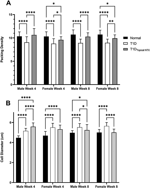
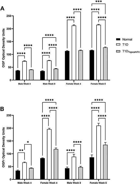
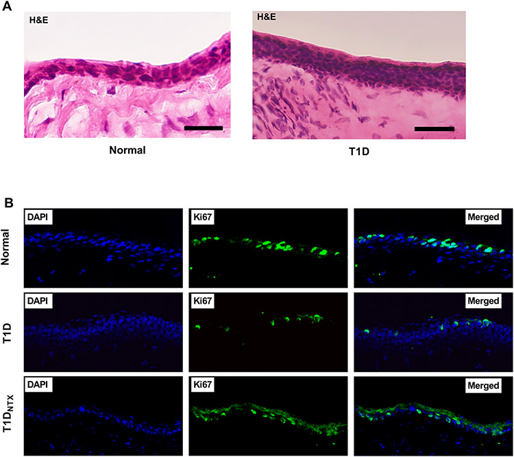
Ocular surface complications occur in more than 50% of individuals diagnosed with diabetes. The financial and health-related burden of diabetes is increasing annually. Several major ocular complications associated with diabetes involve the limbus. The vascular limbus, adjacent to the avascular cornea, is the source of circulating growth factors, elevated glucose, and cytokines for the cornea. The Opioid Growth Factor (OGF) - Opioid OGF Receptor (OGFr) axis is comprised of its effector peptide, OGF, [Met5]-enkephalin and the nuclear-associated receptor, OGFr, and has been demonstrated to be dysfunctional in diabetes with elevated serum and tissue levels of the inhibitory growth factor OGF recorded in corneal tissue. Little is known regarding the impact of OGF-OGFr axis dysregulation in diabetes on the functioning of the limbus constituents in support of corneal homeostasis. Adult male and female Sprague-Dawley rats were rendered hyperglycemic through intraperitoneal injections of streptozotocin (T1D); a subset of T1D rats received topical naltrexone (NTX) applied to the cornea and limbus daily for 8 weeks. At 4 and/or 8 weeks of hyperglycemia, different cohorts of animals were euthanized, eyes removed and processed for assessment of limbal morphology, expression of OGF, OGFr, cytokeratin 15, a marker for limbal cells, and Ki-67, a marker of proliferation. Limbal epithelial morphology (cell diameter, packing density) was altered in T1D male and female rats. OGF and OGFr were overexpressed in the limbus and CK15 expression was decreased, relative to normal control rats of the same sex. Blockade of the OGF- OGFr axis with NTX reversed limbal epithelial cell defects, and reduced OGF limbal tissue levels to those recorded in non-diabetic rats. In summary, OGF-OGFr axis dysregulation was observed in the limbus of T1D rats, contributing to the altered limbal morphology and delayed corneal surface healing observed in diabetic animals.


 分享
分享

 求助内容:
求助内容: 应助结果提醒方式:
 扫码关注我们
