Duarte Vaz Pimentel, Larissa Merten, Jan-Hendrik Gosemann, Ines Gockel, Boris Jansen-Winkeln, Steffi Mayer, Martin Lacher
{"title":"高光谱成像-一种评估食管吻合口组织灌注和氧合的新工具。","authors":"Duarte Vaz Pimentel, Larissa Merten, Jan-Hendrik Gosemann, Ines Gockel, Boris Jansen-Winkeln, Steffi Mayer, Martin Lacher","doi":"10.1055/s-0043-1769106","DOIUrl":null,"url":null,"abstract":"<p><p>Anastomotic stricture and leakage are common complications after repair of esophageal atresia (EA). A compromised perfusion of the anastomosis is a contributing factor. Hyperspectral imaging (HSI) is an ultrashort noninvasive method to measure tissue perfusion. We present two cases of with tracheoesophageal fistula (TEF)/EA repair, in whom we applied HSI: the first patient was a newborn with EA type C who underwent open TEF repair. The second one had an EA type A and cervical esophagostomy, in whom we performed gastric transposition. In both patients, HSI confirmed a good tissue perfusion of the later anastomosis. The postoperative course was uneventful and both patients are on full enteral feeds. We conclude that HSI is a safe and noninvasive tool that allows near real-time assessment of tissue perfusion and can contribute to the identification of the optimal anastomotic region during pediatric esophageal surgery.</p>","PeriodicalId":43204,"journal":{"name":"European Journal of Pediatric Surgery Reports","volume":"11 1","pages":"e32-e35"},"PeriodicalIF":0.7000,"publicationDate":"2023-01-01","publicationTypes":"Journal Article","fieldsOfStudy":null,"isOpenAccess":false,"openAccessPdf":"https://www.ncbi.nlm.nih.gov/pmc/articles/PMC10260350/pdf/","citationCount":"0","resultStr":"{\"title\":\"Hyperspectral Imaging-A Novel Tool to Assess Tissue Perfusion and Oxygenation in Esophageal Anastomoses.\",\"authors\":\"Duarte Vaz Pimentel, Larissa Merten, Jan-Hendrik Gosemann, Ines Gockel, Boris Jansen-Winkeln, Steffi Mayer, Martin Lacher\",\"doi\":\"10.1055/s-0043-1769106\",\"DOIUrl\":null,\"url\":null,\"abstract\":\"<p><p>Anastomotic stricture and leakage are common complications after repair of esophageal atresia (EA). A compromised perfusion of the anastomosis is a contributing factor. Hyperspectral imaging (HSI) is an ultrashort noninvasive method to measure tissue perfusion. We present two cases of with tracheoesophageal fistula (TEF)/EA repair, in whom we applied HSI: the first patient was a newborn with EA type C who underwent open TEF repair. The second one had an EA type A and cervical esophagostomy, in whom we performed gastric transposition. In both patients, HSI confirmed a good tissue perfusion of the later anastomosis. The postoperative course was uneventful and both patients are on full enteral feeds. We conclude that HSI is a safe and noninvasive tool that allows near real-time assessment of tissue perfusion and can contribute to the identification of the optimal anastomotic region during pediatric esophageal surgery.</p>\",\"PeriodicalId\":43204,\"journal\":{\"name\":\"European Journal of Pediatric Surgery Reports\",\"volume\":\"11 1\",\"pages\":\"e32-e35\"},\"PeriodicalIF\":0.7000,\"publicationDate\":\"2023-01-01\",\"publicationTypes\":\"Journal Article\",\"fieldsOfStudy\":null,\"isOpenAccess\":false,\"openAccessPdf\":\"https://www.ncbi.nlm.nih.gov/pmc/articles/PMC10260350/pdf/\",\"citationCount\":\"0\",\"resultStr\":null,\"platform\":\"Semanticscholar\",\"paperid\":null,\"PeriodicalName\":\"European Journal of Pediatric Surgery Reports\",\"FirstCategoryId\":\"1085\",\"ListUrlMain\":\"https://doi.org/10.1055/s-0043-1769106\",\"RegionNum\":0,\"RegionCategory\":null,\"ArticlePicture\":[],\"TitleCN\":null,\"AbstractTextCN\":null,\"PMCID\":null,\"EPubDate\":\"\",\"PubModel\":\"\",\"JCR\":\"Q4\",\"JCRName\":\"SURGERY\",\"Score\":null,\"Total\":0}","platform":"Semanticscholar","paperid":null,"PeriodicalName":"European Journal of Pediatric Surgery Reports","FirstCategoryId":"1085","ListUrlMain":"https://doi.org/10.1055/s-0043-1769106","RegionNum":0,"RegionCategory":null,"ArticlePicture":[],"TitleCN":null,"AbstractTextCN":null,"PMCID":null,"EPubDate":"","PubModel":"","JCR":"Q4","JCRName":"SURGERY","Score":null,"Total":0}
Hyperspectral Imaging-A Novel Tool to Assess Tissue Perfusion and Oxygenation in Esophageal Anastomoses.
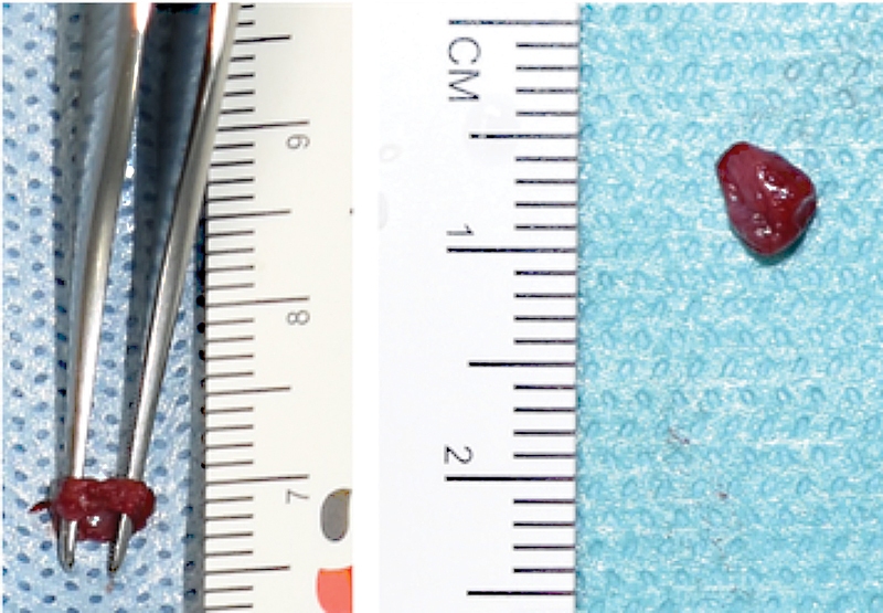
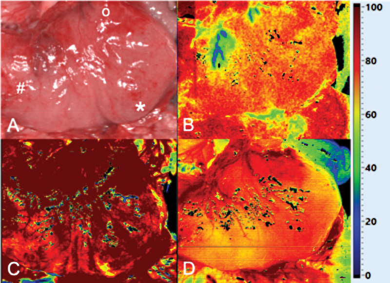
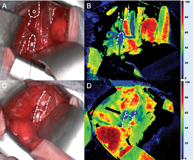
Anastomotic stricture and leakage are common complications after repair of esophageal atresia (EA). A compromised perfusion of the anastomosis is a contributing factor. Hyperspectral imaging (HSI) is an ultrashort noninvasive method to measure tissue perfusion. We present two cases of with tracheoesophageal fistula (TEF)/EA repair, in whom we applied HSI: the first patient was a newborn with EA type C who underwent open TEF repair. The second one had an EA type A and cervical esophagostomy, in whom we performed gastric transposition. In both patients, HSI confirmed a good tissue perfusion of the later anastomosis. The postoperative course was uneventful and both patients are on full enteral feeds. We conclude that HSI is a safe and noninvasive tool that allows near real-time assessment of tissue perfusion and can contribute to the identification of the optimal anastomotic region during pediatric esophageal surgery.


 分享
分享

 求助内容:
求助内容: 应助结果提醒方式:
 扫码关注我们
