{"title":"Myomaker和mymerger在肌纤维中的表达引起肌肉病理。","authors":"Phillip C Witcher, Chengyi Sun, Douglas P Millay","doi":"10.1186/s13395-023-00317-z","DOIUrl":null,"url":null,"abstract":"<p><strong>Background: </strong>Skeletal muscle development and regeneration depend on cellular fusion of myogenic progenitors to generate multinucleated myofibers. These progenitors utilize two muscle-specific fusogens, Myomaker and Myomerger, which function by remodeling cell membranes to fuse to each other or to existing myofibers. Myomaker and Myomerger expression is restricted to differentiating progenitor cells as they are not detected in adult myofibers. However, Myomaker remains expressed in myofibers from mice with muscular dystrophy. Ablation of Myomaker from dystrophic myofibers results in reduced membrane damage, leading to a model where persistent fusogen expression in myofibers, in contrast to myoblasts, is harmful.</p><p><strong>Methods: </strong>Dox-inducible transgenic mice were developed to ectopically express Myomaker or Myomerger in the myofiber compartment of skeletal muscle. We quantified indices of myofiber membrane damage, such as serum creatine kinase and IgM<sup>+</sup> myofibers, and assessed general muscle histology, including central nucleation, myofiber size, and fibrosis.</p><p><strong>Results: </strong>Myomaker or Myomerger expression in myofibers independently caused membrane damage at acute time points. This damage led to muscle pathology, manifesting with centrally nucleated myofibers and muscle atrophy. Dual expression of both Myomaker and Myomerger in myofibers exacerbated several aspects of muscle pathology compared to expression of either fusogen by itself.</p><p><strong>Conclusions: </strong>These data reveal that while myofibers can tolerate some level of Myomaker and Myomerger, expression of a single fusogen above a threshold or co-expression of both fusogens is damaging to myofibers. These results explain the paradigm that their expression in myofibers can have deleterious consequences in muscle pathologies and highlight the need for their highly restricted expression during myogenesis and fusion.</p>","PeriodicalId":21747,"journal":{"name":"Skeletal Muscle","volume":"13 1","pages":"8"},"PeriodicalIF":4.4000,"publicationDate":"2023-05-01","publicationTypes":"Journal Article","fieldsOfStudy":null,"isOpenAccess":false,"openAccessPdf":"https://www.ncbi.nlm.nih.gov/pmc/articles/PMC10150476/pdf/","citationCount":"1","resultStr":"{\"title\":\"Expression of Myomaker and Myomerger in myofibers causes muscle pathology.\",\"authors\":\"Phillip C Witcher, Chengyi Sun, Douglas P Millay\",\"doi\":\"10.1186/s13395-023-00317-z\",\"DOIUrl\":null,\"url\":null,\"abstract\":\"<p><strong>Background: </strong>Skeletal muscle development and regeneration depend on cellular fusion of myogenic progenitors to generate multinucleated myofibers. These progenitors utilize two muscle-specific fusogens, Myomaker and Myomerger, which function by remodeling cell membranes to fuse to each other or to existing myofibers. Myomaker and Myomerger expression is restricted to differentiating progenitor cells as they are not detected in adult myofibers. However, Myomaker remains expressed in myofibers from mice with muscular dystrophy. Ablation of Myomaker from dystrophic myofibers results in reduced membrane damage, leading to a model where persistent fusogen expression in myofibers, in contrast to myoblasts, is harmful.</p><p><strong>Methods: </strong>Dox-inducible transgenic mice were developed to ectopically express Myomaker or Myomerger in the myofiber compartment of skeletal muscle. We quantified indices of myofiber membrane damage, such as serum creatine kinase and IgM<sup>+</sup> myofibers, and assessed general muscle histology, including central nucleation, myofiber size, and fibrosis.</p><p><strong>Results: </strong>Myomaker or Myomerger expression in myofibers independently caused membrane damage at acute time points. This damage led to muscle pathology, manifesting with centrally nucleated myofibers and muscle atrophy. Dual expression of both Myomaker and Myomerger in myofibers exacerbated several aspects of muscle pathology compared to expression of either fusogen by itself.</p><p><strong>Conclusions: </strong>These data reveal that while myofibers can tolerate some level of Myomaker and Myomerger, expression of a single fusogen above a threshold or co-expression of both fusogens is damaging to myofibers. These results explain the paradigm that their expression in myofibers can have deleterious consequences in muscle pathologies and highlight the need for their highly restricted expression during myogenesis and fusion.</p>\",\"PeriodicalId\":21747,\"journal\":{\"name\":\"Skeletal Muscle\",\"volume\":\"13 1\",\"pages\":\"8\"},\"PeriodicalIF\":4.4000,\"publicationDate\":\"2023-05-01\",\"publicationTypes\":\"Journal Article\",\"fieldsOfStudy\":null,\"isOpenAccess\":false,\"openAccessPdf\":\"https://www.ncbi.nlm.nih.gov/pmc/articles/PMC10150476/pdf/\",\"citationCount\":\"1\",\"resultStr\":null,\"platform\":\"Semanticscholar\",\"paperid\":null,\"PeriodicalName\":\"Skeletal Muscle\",\"FirstCategoryId\":\"3\",\"ListUrlMain\":\"https://doi.org/10.1186/s13395-023-00317-z\",\"RegionNum\":2,\"RegionCategory\":\"医学\",\"ArticlePicture\":[],\"TitleCN\":null,\"AbstractTextCN\":null,\"PMCID\":null,\"EPubDate\":\"\",\"PubModel\":\"\",\"JCR\":\"Q2\",\"JCRName\":\"CELL BIOLOGY\",\"Score\":null,\"Total\":0}","platform":"Semanticscholar","paperid":null,"PeriodicalName":"Skeletal Muscle","FirstCategoryId":"3","ListUrlMain":"https://doi.org/10.1186/s13395-023-00317-z","RegionNum":2,"RegionCategory":"医学","ArticlePicture":[],"TitleCN":null,"AbstractTextCN":null,"PMCID":null,"EPubDate":"","PubModel":"","JCR":"Q2","JCRName":"CELL BIOLOGY","Score":null,"Total":0}
Expression of Myomaker and Myomerger in myofibers causes muscle pathology.
Background: Skeletal muscle development and regeneration depend on cellular fusion of myogenic progenitors to generate multinucleated myofibers. These progenitors utilize two muscle-specific fusogens, Myomaker and Myomerger, which function by remodeling cell membranes to fuse to each other or to existing myofibers. Myomaker and Myomerger expression is restricted to differentiating progenitor cells as they are not detected in adult myofibers. However, Myomaker remains expressed in myofibers from mice with muscular dystrophy. Ablation of Myomaker from dystrophic myofibers results in reduced membrane damage, leading to a model where persistent fusogen expression in myofibers, in contrast to myoblasts, is harmful.
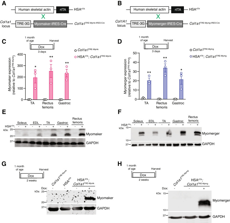
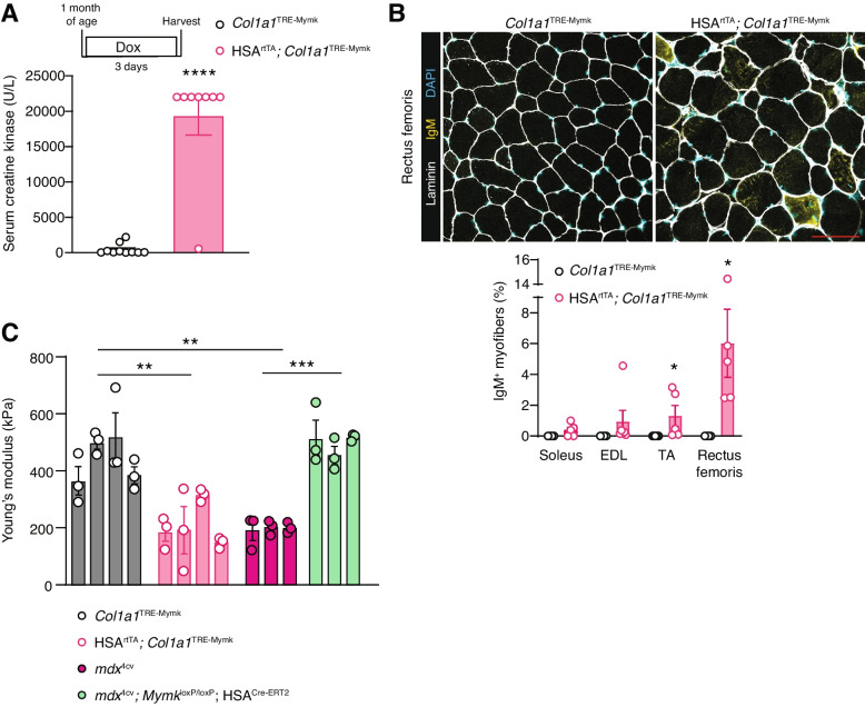
Methods: Dox-inducible transgenic mice were developed to ectopically express Myomaker or Myomerger in the myofiber compartment of skeletal muscle. We quantified indices of myofiber membrane damage, such as serum creatine kinase and IgM+ myofibers, and assessed general muscle histology, including central nucleation, myofiber size, and fibrosis.
Results: Myomaker or Myomerger expression in myofibers independently caused membrane damage at acute time points. This damage led to muscle pathology, manifesting with centrally nucleated myofibers and muscle atrophy. Dual expression of both Myomaker and Myomerger in myofibers exacerbated several aspects of muscle pathology compared to expression of either fusogen by itself.
Conclusions: These data reveal that while myofibers can tolerate some level of Myomaker and Myomerger, expression of a single fusogen above a threshold or co-expression of both fusogens is damaging to myofibers. These results explain the paradigm that their expression in myofibers can have deleterious consequences in muscle pathologies and highlight the need for their highly restricted expression during myogenesis and fusion.
期刊介绍:
The only open access journal in its field, Skeletal Muscle publishes novel, cutting-edge research and technological advancements that investigate the molecular mechanisms underlying the biology of skeletal muscle. Reflecting the breadth of research in this area, the journal welcomes manuscripts about the development, metabolism, the regulation of mass and function, aging, degeneration, dystrophy and regeneration of skeletal muscle, with an emphasis on understanding adult skeletal muscle, its maintenance, and its interactions with non-muscle cell types and regulatory modulators.
Main areas of interest include:
-differentiation of skeletal muscle-
atrophy and hypertrophy of skeletal muscle-
aging of skeletal muscle-
regeneration and degeneration of skeletal muscle-
biology of satellite and satellite-like cells-
dystrophic degeneration of skeletal muscle-
energy and glucose homeostasis in skeletal muscle-
non-dystrophic genetic diseases of skeletal muscle, such as Spinal Muscular Atrophy and myopathies-
maintenance of neuromuscular junctions-
roles of ryanodine receptors and calcium signaling in skeletal muscle-
roles of nuclear receptors in skeletal muscle-
roles of GPCRs and GPCR signaling in skeletal muscle-
other relevant aspects of skeletal muscle biology.
In addition, articles on translational clinical studies that address molecular and cellular mechanisms of skeletal muscle will be published. Case reports are also encouraged for submission.
Skeletal Muscle reflects the breadth of research on skeletal muscle and bridges gaps between diverse areas of science for example cardiac cell biology and neurobiology, which share common features with respect to cell differentiation, excitatory membranes, cell-cell communication, and maintenance. Suitable articles are model and mechanism-driven, and apply statistical principles where appropriate; purely descriptive studies are of lesser interest.


 分享
分享
 求助内容:
求助内容: 应助结果提醒方式:
 扫码关注我们
