Sheng Cheng, Jutang Li, Ming Xu, Qun Bao, Jiaoxiang Wu, Peng Sun, Bo Han
{"title":"TMEM147与免疫浸润相关,可作为肝细胞癌的潜在预后生物标志物","authors":"Sheng Cheng, Jutang Li, Ming Xu, Qun Bao, Jiaoxiang Wu, Peng Sun, Bo Han","doi":"10.1155/2023/4413049","DOIUrl":null,"url":null,"abstract":"<p><strong>Background: </strong>Hepatocellular carcinoma (HCC) is one of the most prevalent malignancies and is associated with high mortality. Transmembrane protein 147 (TMEM147) is a seven-transmembrane protein that may mediate immune regulation. However, the relevance of TMEM147 to immune regulation in HCC and the prognosis of HCC patients are unclear.</p><p><strong>Methods: </strong>We analyzed TMEM147 expression in HCC by using the Wilcoxon rank-sum test. Real time quantitative PCR (RT-qPCR) and Western blot analysis of tumor tissues and cell lines were used to verify TMEM147 expression in HCC. The influence of TMEM147 on HCC prognosis was assessed using Kaplan-Meier analysis, Cox regression analysis, and a prognostic nomogram. The functions of the TMEM147-related differentially expressed genes (DEGs) were identified by Gene Ontology (GO)/Kyoto Encyclopedia of Genes and Genomes (KEGG) enrichment analyses and gene set enrichment analysis (GSEA). In addition, we examined the associations between TMEM147 expression and immune infiltration using single-sample gene set enrichment analysis (ssGSEA) and immunofluorescence staining of HCC tissues.</p><p><strong>Results: </strong>Our results showed that the expression of TMEM147 was significantly higher in human HCC tissues than in adjacent normal liver tissues, with similar findings in human HCC cell lines. High TMEM147 expression was correlated with T stage, pathological stage, histological grade, race, alpha-fetoprotein level, and vascular invasion in HCC. Moreover, we revealed that high TMEM147 expression was associated with shorter survival times and that TMEM147 could be a risk factor for overall survival, along with T stage, M stage, pathological stage, and tumor status. Mechanistic studies revealed that high TMEM147 expression was linked to the B lymphocyte, antigen response, IL6 signaling pathway, cell cycle, Kirsten rat sarcoma viral oncogene homolog (KRAS) signaling pathway, and myelocytomatosis oncogene (MYC) targets. Correspondingly, TMEM147 expression was positively associated with the infiltration of immune cells, including Th2 cells, follicular helper T cells, macrophages, and NK CD56 bright cells in HCC.</p><p><strong>Conclusions: </strong>TMEM147 might be a biomarker for poor prognosis and is related to immune cell infiltration in HCC.</p>","PeriodicalId":49326,"journal":{"name":"Analytical Cellular Pathology","volume":"2023 ","pages":"4413049"},"PeriodicalIF":2.7000,"publicationDate":"2023-01-01","publicationTypes":"Journal Article","fieldsOfStudy":null,"isOpenAccess":false,"openAccessPdf":"https://www.ncbi.nlm.nih.gov/pmc/articles/PMC10257544/pdf/","citationCount":"0","resultStr":"{\"title\":\"TMEM147 Correlates with Immune Infiltration and Serve as a Potential Prognostic Biomarker in Hepatocellular Carcinoma.\",\"authors\":\"Sheng Cheng, Jutang Li, Ming Xu, Qun Bao, Jiaoxiang Wu, Peng Sun, Bo Han\",\"doi\":\"10.1155/2023/4413049\",\"DOIUrl\":null,\"url\":null,\"abstract\":\"<p><strong>Background: </strong>Hepatocellular carcinoma (HCC) is one of the most prevalent malignancies and is associated with high mortality. Transmembrane protein 147 (TMEM147) is a seven-transmembrane protein that may mediate immune regulation. However, the relevance of TMEM147 to immune regulation in HCC and the prognosis of HCC patients are unclear.</p><p><strong>Methods: </strong>We analyzed TMEM147 expression in HCC by using the Wilcoxon rank-sum test. Real time quantitative PCR (RT-qPCR) and Western blot analysis of tumor tissues and cell lines were used to verify TMEM147 expression in HCC. The influence of TMEM147 on HCC prognosis was assessed using Kaplan-Meier analysis, Cox regression analysis, and a prognostic nomogram. The functions of the TMEM147-related differentially expressed genes (DEGs) were identified by Gene Ontology (GO)/Kyoto Encyclopedia of Genes and Genomes (KEGG) enrichment analyses and gene set enrichment analysis (GSEA). In addition, we examined the associations between TMEM147 expression and immune infiltration using single-sample gene set enrichment analysis (ssGSEA) and immunofluorescence staining of HCC tissues.</p><p><strong>Results: </strong>Our results showed that the expression of TMEM147 was significantly higher in human HCC tissues than in adjacent normal liver tissues, with similar findings in human HCC cell lines. High TMEM147 expression was correlated with T stage, pathological stage, histological grade, race, alpha-fetoprotein level, and vascular invasion in HCC. Moreover, we revealed that high TMEM147 expression was associated with shorter survival times and that TMEM147 could be a risk factor for overall survival, along with T stage, M stage, pathological stage, and tumor status. Mechanistic studies revealed that high TMEM147 expression was linked to the B lymphocyte, antigen response, IL6 signaling pathway, cell cycle, Kirsten rat sarcoma viral oncogene homolog (KRAS) signaling pathway, and myelocytomatosis oncogene (MYC) targets. Correspondingly, TMEM147 expression was positively associated with the infiltration of immune cells, including Th2 cells, follicular helper T cells, macrophages, and NK CD56 bright cells in HCC.</p><p><strong>Conclusions: </strong>TMEM147 might be a biomarker for poor prognosis and is related to immune cell infiltration in HCC.</p>\",\"PeriodicalId\":49326,\"journal\":{\"name\":\"Analytical Cellular Pathology\",\"volume\":\"2023 \",\"pages\":\"4413049\"},\"PeriodicalIF\":2.7000,\"publicationDate\":\"2023-01-01\",\"publicationTypes\":\"Journal Article\",\"fieldsOfStudy\":null,\"isOpenAccess\":false,\"openAccessPdf\":\"https://www.ncbi.nlm.nih.gov/pmc/articles/PMC10257544/pdf/\",\"citationCount\":\"0\",\"resultStr\":null,\"platform\":\"Semanticscholar\",\"paperid\":null,\"PeriodicalName\":\"Analytical Cellular Pathology\",\"FirstCategoryId\":\"3\",\"ListUrlMain\":\"https://doi.org/10.1155/2023/4413049\",\"RegionNum\":4,\"RegionCategory\":\"医学\",\"ArticlePicture\":[],\"TitleCN\":null,\"AbstractTextCN\":null,\"PMCID\":null,\"EPubDate\":\"\",\"PubModel\":\"\",\"JCR\":\"Q3\",\"JCRName\":\"CELL BIOLOGY\",\"Score\":null,\"Total\":0}","platform":"Semanticscholar","paperid":null,"PeriodicalName":"Analytical Cellular Pathology","FirstCategoryId":"3","ListUrlMain":"https://doi.org/10.1155/2023/4413049","RegionNum":4,"RegionCategory":"医学","ArticlePicture":[],"TitleCN":null,"AbstractTextCN":null,"PMCID":null,"EPubDate":"","PubModel":"","JCR":"Q3","JCRName":"CELL BIOLOGY","Score":null,"Total":0}
TMEM147 Correlates with Immune Infiltration and Serve as a Potential Prognostic Biomarker in Hepatocellular Carcinoma.
Background: Hepatocellular carcinoma (HCC) is one of the most prevalent malignancies and is associated with high mortality. Transmembrane protein 147 (TMEM147) is a seven-transmembrane protein that may mediate immune regulation. However, the relevance of TMEM147 to immune regulation in HCC and the prognosis of HCC patients are unclear.
Methods: We analyzed TMEM147 expression in HCC by using the Wilcoxon rank-sum test. Real time quantitative PCR (RT-qPCR) and Western blot analysis of tumor tissues and cell lines were used to verify TMEM147 expression in HCC. The influence of TMEM147 on HCC prognosis was assessed using Kaplan-Meier analysis, Cox regression analysis, and a prognostic nomogram. The functions of the TMEM147-related differentially expressed genes (DEGs) were identified by Gene Ontology (GO)/Kyoto Encyclopedia of Genes and Genomes (KEGG) enrichment analyses and gene set enrichment analysis (GSEA). In addition, we examined the associations between TMEM147 expression and immune infiltration using single-sample gene set enrichment analysis (ssGSEA) and immunofluorescence staining of HCC tissues.
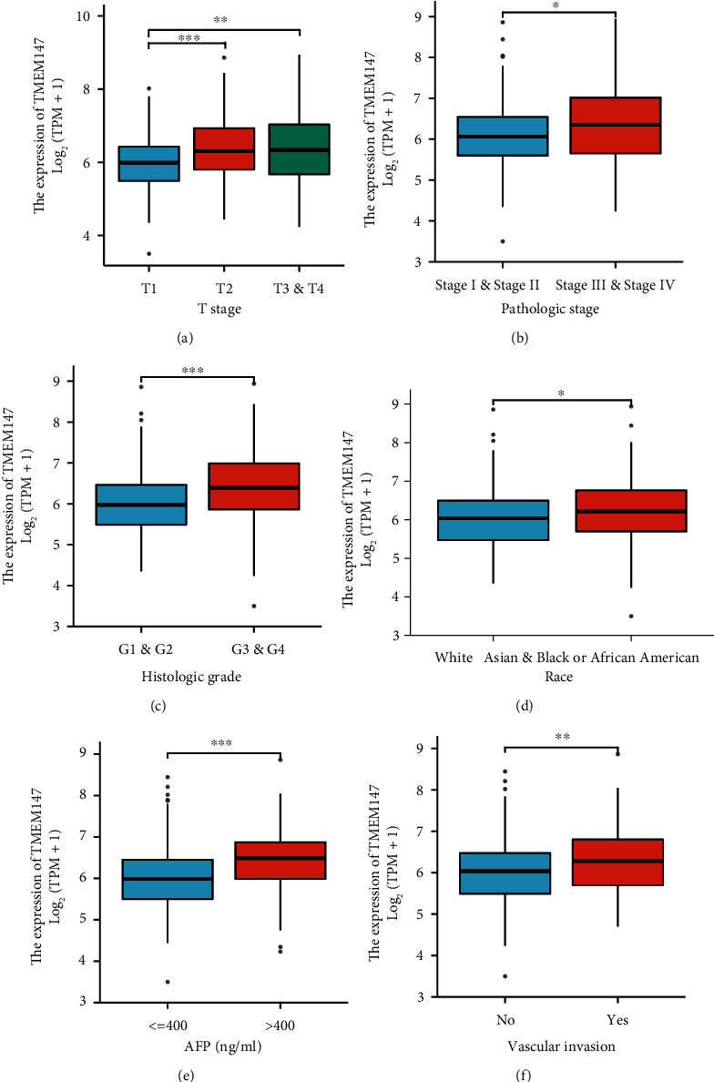
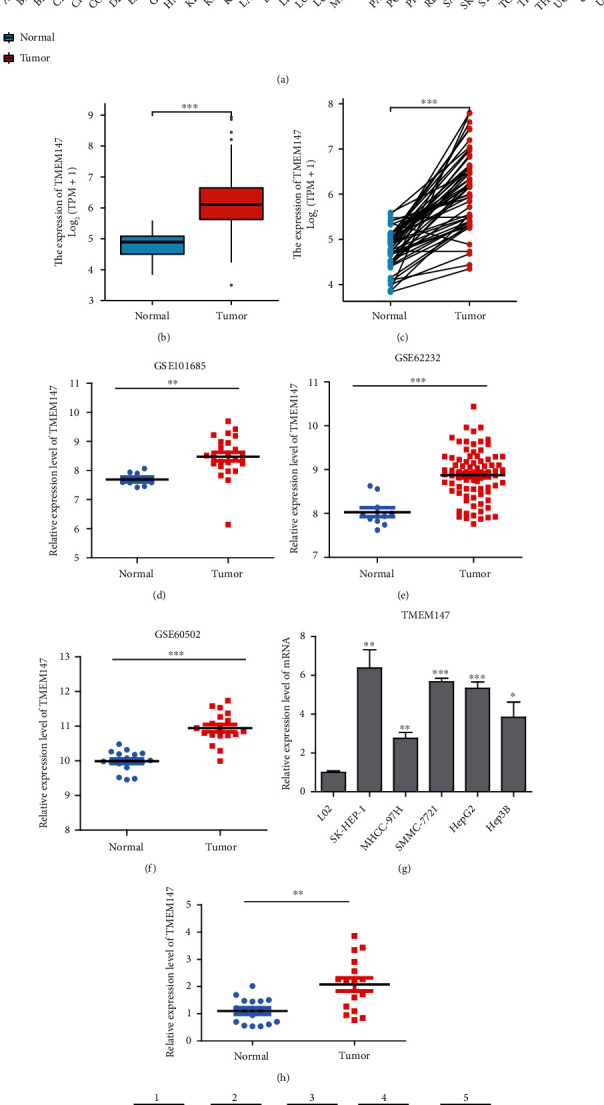
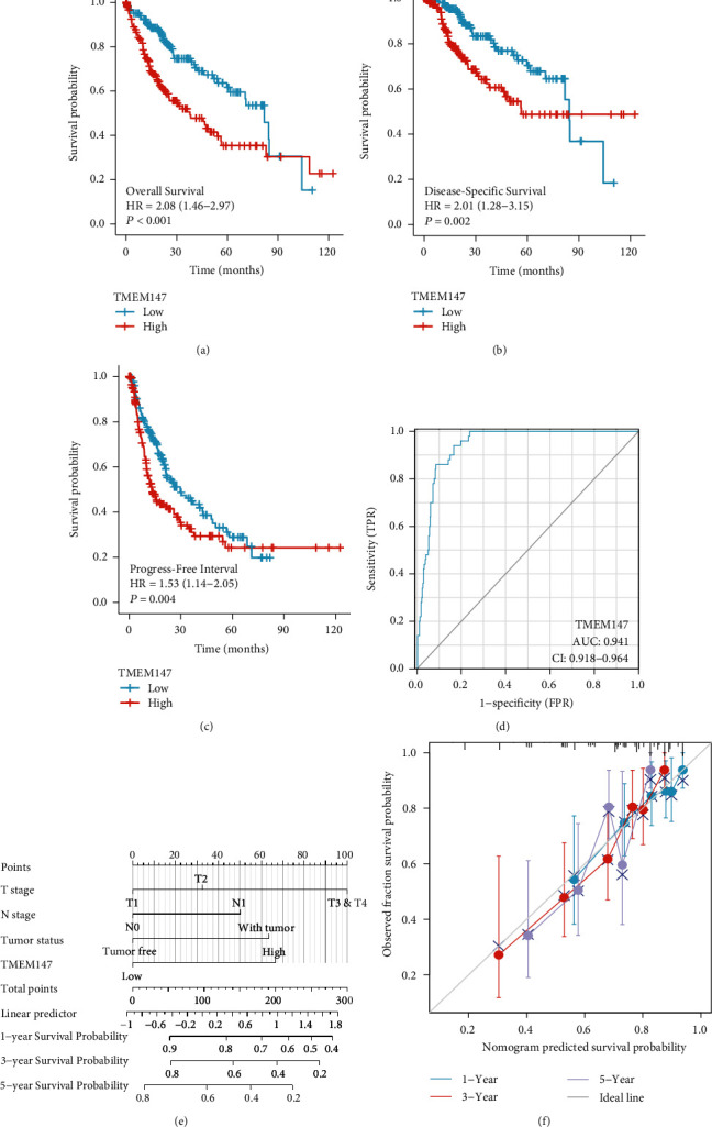
Results: Our results showed that the expression of TMEM147 was significantly higher in human HCC tissues than in adjacent normal liver tissues, with similar findings in human HCC cell lines. High TMEM147 expression was correlated with T stage, pathological stage, histological grade, race, alpha-fetoprotein level, and vascular invasion in HCC. Moreover, we revealed that high TMEM147 expression was associated with shorter survival times and that TMEM147 could be a risk factor for overall survival, along with T stage, M stage, pathological stage, and tumor status. Mechanistic studies revealed that high TMEM147 expression was linked to the B lymphocyte, antigen response, IL6 signaling pathway, cell cycle, Kirsten rat sarcoma viral oncogene homolog (KRAS) signaling pathway, and myelocytomatosis oncogene (MYC) targets. Correspondingly, TMEM147 expression was positively associated with the infiltration of immune cells, including Th2 cells, follicular helper T cells, macrophages, and NK CD56 bright cells in HCC.
Conclusions: TMEM147 might be a biomarker for poor prognosis and is related to immune cell infiltration in HCC.
期刊介绍:
Analytical Cellular Pathology is a peer-reviewed, Open Access journal that provides a forum for scientists, medical practitioners and pathologists working in the area of cellular pathology. The journal publishes original research articles, review articles, and clinical studies related to cytology, carcinogenesis, cell receptors, biomarkers, diagnostic pathology, immunopathology, and hematology.


 分享
分享
 求助内容:
求助内容: 应助结果提醒方式:
 扫码关注我们
