{"title":"[PI-RADS 2.1和前列腺磁共振成像的结构化报告]。","authors":"Andreas Hötker, Olivio F Donati","doi":"10.1007/s00117-021-00868-6","DOIUrl":null,"url":null,"abstract":"<p><strong>Clinical/methodological issue: </strong>The detection of clinically significant prostate cancers while simultaneously avoiding over-diagnosing tumors with low malignant potential is a challenge in clinical practice.</p><p><strong>Standard radiological methods: </strong>Multiparametric prostate magnetic resonance imaging (MRI) in accordance with the Prostate Imaging Reporting and Data System (PI-RADS) guidelines is accepted as standard-of-care with both urologists and radiologists.</p><p><strong>Methodological innovations: </strong>The PI-RADS guidelines have been updated to version 2.1, including revised technical recommendations and changes to the scoring of lesions.</p><p><strong>Performance: </strong>The PI-RADS guidelines have had great impact on the standardization of multiparametric prostate MRI and offer templates for structured reporting. This simplifies communication with the referring physician.</p><p><strong>Achievements: </strong>The new version 2.1 of the guidelines represents an evolutionary improvement of the widely accepted version 2.0. Several aspects of reporting have been revised-however, some pre-known limitations persist, which will require further refinement in the future.</p>","PeriodicalId":54513,"journal":{"name":"Radiologe","volume":"61 9","pages":"802-809"},"PeriodicalIF":0.0000,"publicationDate":"2021-09-01","publicationTypes":"Journal Article","fieldsOfStudy":null,"isOpenAccess":false,"openAccessPdf":"https://sci-hub-pdf.com/10.1007/s00117-021-00868-6","citationCount":"2","resultStr":"{\"title\":\"[PI-RADS 2.1 and structured reporting of magnetic resonance imaging of the prostate].\",\"authors\":\"Andreas Hötker, Olivio F Donati\",\"doi\":\"10.1007/s00117-021-00868-6\",\"DOIUrl\":null,\"url\":null,\"abstract\":\"<p><strong>Clinical/methodological issue: </strong>The detection of clinically significant prostate cancers while simultaneously avoiding over-diagnosing tumors with low malignant potential is a challenge in clinical practice.</p><p><strong>Standard radiological methods: </strong>Multiparametric prostate magnetic resonance imaging (MRI) in accordance with the Prostate Imaging Reporting and Data System (PI-RADS) guidelines is accepted as standard-of-care with both urologists and radiologists.</p><p><strong>Methodological innovations: </strong>The PI-RADS guidelines have been updated to version 2.1, including revised technical recommendations and changes to the scoring of lesions.</p><p><strong>Performance: </strong>The PI-RADS guidelines have had great impact on the standardization of multiparametric prostate MRI and offer templates for structured reporting. This simplifies communication with the referring physician.</p><p><strong>Achievements: </strong>The new version 2.1 of the guidelines represents an evolutionary improvement of the widely accepted version 2.0. Several aspects of reporting have been revised-however, some pre-known limitations persist, which will require further refinement in the future.</p>\",\"PeriodicalId\":54513,\"journal\":{\"name\":\"Radiologe\",\"volume\":\"61 9\",\"pages\":\"802-809\"},\"PeriodicalIF\":0.0000,\"publicationDate\":\"2021-09-01\",\"publicationTypes\":\"Journal Article\",\"fieldsOfStudy\":null,\"isOpenAccess\":false,\"openAccessPdf\":\"https://sci-hub-pdf.com/10.1007/s00117-021-00868-6\",\"citationCount\":\"2\",\"resultStr\":null,\"platform\":\"Semanticscholar\",\"paperid\":null,\"PeriodicalName\":\"Radiologe\",\"FirstCategoryId\":\"3\",\"ListUrlMain\":\"https://doi.org/10.1007/s00117-021-00868-6\",\"RegionNum\":4,\"RegionCategory\":\"医学\",\"ArticlePicture\":[],\"TitleCN\":null,\"AbstractTextCN\":null,\"PMCID\":null,\"EPubDate\":\"\",\"PubModel\":\"\",\"JCR\":\"Q3\",\"JCRName\":\"Medicine\",\"Score\":null,\"Total\":0}","platform":"Semanticscholar","paperid":null,"PeriodicalName":"Radiologe","FirstCategoryId":"3","ListUrlMain":"https://doi.org/10.1007/s00117-021-00868-6","RegionNum":4,"RegionCategory":"医学","ArticlePicture":[],"TitleCN":null,"AbstractTextCN":null,"PMCID":null,"EPubDate":"","PubModel":"","JCR":"Q3","JCRName":"Medicine","Score":null,"Total":0}
[PI-RADS 2.1 and structured reporting of magnetic resonance imaging of the prostate].
Clinical/methodological issue: The detection of clinically significant prostate cancers while simultaneously avoiding over-diagnosing tumors with low malignant potential is a challenge in clinical practice.
Standard radiological methods: Multiparametric prostate magnetic resonance imaging (MRI) in accordance with the Prostate Imaging Reporting and Data System (PI-RADS) guidelines is accepted as standard-of-care with both urologists and radiologists.
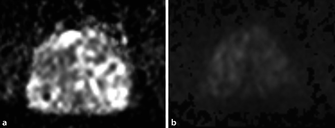
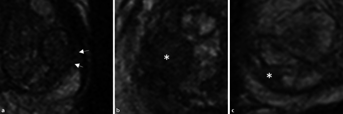
Methodological innovations: The PI-RADS guidelines have been updated to version 2.1, including revised technical recommendations and changes to the scoring of lesions.
Performance: The PI-RADS guidelines have had great impact on the standardization of multiparametric prostate MRI and offer templates for structured reporting. This simplifies communication with the referring physician.
Achievements: The new version 2.1 of the guidelines represents an evolutionary improvement of the widely accepted version 2.0. Several aspects of reporting have been revised-however, some pre-known limitations persist, which will require further refinement in the future.
期刊介绍:
Der Radiologe is an internationally recognized journal dealing with all aspects of radiology and serving the continuing medical education of radiologists in clinical and practical environments. The focus is on x-ray diagnostics, angiography computer tomography, interventional radiology, magnet resonance tomography, digital picture processing, radio oncology and nuclear medicine.
Comprehensive reviews on a specific topical issue focus on providing evidenced based information on diagnostics and therapy.
Freely submitted original papers allow the presentation of important clinical studies and serve the scientific exchange.
Review articles under the rubric ''Continuing Medical Education'' present verified results of scientific research and their integration into daily practice.


 分享
分享
 求助内容:
求助内容: 应助结果提醒方式:
 扫码关注我们
