Rita Castro, Sean Gullette, Courtney Whalen, Floyd J Mattie, Ximing Ge, A Catharine Ross, Thomas Neuberger
{"title":"小鼠动脉粥样硬化模型主动脉斑块的高场磁共振显微镜观察。","authors":"Rita Castro, Sean Gullette, Courtney Whalen, Floyd J Mattie, Ximing Ge, A Catharine Ross, Thomas Neuberger","doi":"10.1007/s10334-023-01102-1","DOIUrl":null,"url":null,"abstract":"<p><strong>Objectives: </strong>Pre-clinical models of human atherosclerosis are extensively used; however, traditional histological methods do not allow for a holistic view of vascular lesions. We describe an ex-vivo, high-resolution MRI method that allows the 3 dimensional imaging of the vessel for aortic plaque visualization and quantification.</p><p><strong>Materials and methods: </strong>Aortas from apolipoprotein-E-deficient (apoE<sup>-/-</sup>) mice fed an atherogenic diet (group 1) or a control diet (group 2) were subjected to 14 T MR imaging using a 3D gradient echo sequence. The obtained data sets were reconstructed (Matlab), segmented, and analyzed (Avizo). The aortas were further sectioned and subjected to traditional histological analysis (Oil-Red O and hematoxylin staining) for comparison.</p><p><strong>Results: </strong>A resolution up to 15 × 10x10 μm<sup>3</sup> revealed that plaque burden (mm<sup>3</sup>) was significantly (p < 0.05) higher in group 1 (0.41 ± 0.25, n = 4) than in group 2 (0.01 ± 0.01, n = 3). The achieved resolution provided similar detail on the plaque and the vessel wall morphology compared with histology. Digital image segmentation of the aorta's lumen, plaque, and wall offered three-dimensional visualizations of the entire, intact aortas.</p><p><strong>Discussion: </strong>14 T MR microscopy provided histology-like details of pathologically relevant vascular lesions. This work may provide the path research needs to take to enable plaque characterization in clinical applications.</p>","PeriodicalId":18067,"journal":{"name":"Magnetic Resonance Materials in Physics, Biology and Medicine","volume":null,"pages":null},"PeriodicalIF":2.0000,"publicationDate":"2023-12-01","publicationTypes":"Journal Article","fieldsOfStudy":null,"isOpenAccess":false,"openAccessPdf":"https://www.ncbi.nlm.nih.gov/pmc/articles/PMC10667155/pdf/","citationCount":"0","resultStr":"{\"title\":\"High-field magnetic resonance microscopy of aortic plaques in a mouse model of atherosclerosis.\",\"authors\":\"Rita Castro, Sean Gullette, Courtney Whalen, Floyd J Mattie, Ximing Ge, A Catharine Ross, Thomas Neuberger\",\"doi\":\"10.1007/s10334-023-01102-1\",\"DOIUrl\":null,\"url\":null,\"abstract\":\"<p><strong>Objectives: </strong>Pre-clinical models of human atherosclerosis are extensively used; however, traditional histological methods do not allow for a holistic view of vascular lesions. We describe an ex-vivo, high-resolution MRI method that allows the 3 dimensional imaging of the vessel for aortic plaque visualization and quantification.</p><p><strong>Materials and methods: </strong>Aortas from apolipoprotein-E-deficient (apoE<sup>-/-</sup>) mice fed an atherogenic diet (group 1) or a control diet (group 2) were subjected to 14 T MR imaging using a 3D gradient echo sequence. The obtained data sets were reconstructed (Matlab), segmented, and analyzed (Avizo). The aortas were further sectioned and subjected to traditional histological analysis (Oil-Red O and hematoxylin staining) for comparison.</p><p><strong>Results: </strong>A resolution up to 15 × 10x10 μm<sup>3</sup> revealed that plaque burden (mm<sup>3</sup>) was significantly (p < 0.05) higher in group 1 (0.41 ± 0.25, n = 4) than in group 2 (0.01 ± 0.01, n = 3). The achieved resolution provided similar detail on the plaque and the vessel wall morphology compared with histology. Digital image segmentation of the aorta's lumen, plaque, and wall offered three-dimensional visualizations of the entire, intact aortas.</p><p><strong>Discussion: </strong>14 T MR microscopy provided histology-like details of pathologically relevant vascular lesions. This work may provide the path research needs to take to enable plaque characterization in clinical applications.</p>\",\"PeriodicalId\":18067,\"journal\":{\"name\":\"Magnetic Resonance Materials in Physics, Biology and Medicine\",\"volume\":null,\"pages\":null},\"PeriodicalIF\":2.0000,\"publicationDate\":\"2023-12-01\",\"publicationTypes\":\"Journal Article\",\"fieldsOfStudy\":null,\"isOpenAccess\":false,\"openAccessPdf\":\"https://www.ncbi.nlm.nih.gov/pmc/articles/PMC10667155/pdf/\",\"citationCount\":\"0\",\"resultStr\":null,\"platform\":\"Semanticscholar\",\"paperid\":null,\"PeriodicalName\":\"Magnetic Resonance Materials in Physics, Biology and Medicine\",\"FirstCategoryId\":\"3\",\"ListUrlMain\":\"https://doi.org/10.1007/s10334-023-01102-1\",\"RegionNum\":4,\"RegionCategory\":\"医学\",\"ArticlePicture\":[],\"TitleCN\":null,\"AbstractTextCN\":null,\"PMCID\":null,\"EPubDate\":\"2023/7/8 0:00:00\",\"PubModel\":\"Epub\",\"JCR\":\"Q3\",\"JCRName\":\"RADIOLOGY, NUCLEAR MEDICINE & MEDICAL IMAGING\",\"Score\":null,\"Total\":0}","platform":"Semanticscholar","paperid":null,"PeriodicalName":"Magnetic Resonance Materials in Physics, Biology and Medicine","FirstCategoryId":"3","ListUrlMain":"https://doi.org/10.1007/s10334-023-01102-1","RegionNum":4,"RegionCategory":"医学","ArticlePicture":[],"TitleCN":null,"AbstractTextCN":null,"PMCID":null,"EPubDate":"2023/7/8 0:00:00","PubModel":"Epub","JCR":"Q3","JCRName":"RADIOLOGY, NUCLEAR MEDICINE & MEDICAL IMAGING","Score":null,"Total":0}
High-field magnetic resonance microscopy of aortic plaques in a mouse model of atherosclerosis.
Objectives: Pre-clinical models of human atherosclerosis are extensively used; however, traditional histological methods do not allow for a holistic view of vascular lesions. We describe an ex-vivo, high-resolution MRI method that allows the 3 dimensional imaging of the vessel for aortic plaque visualization and quantification.
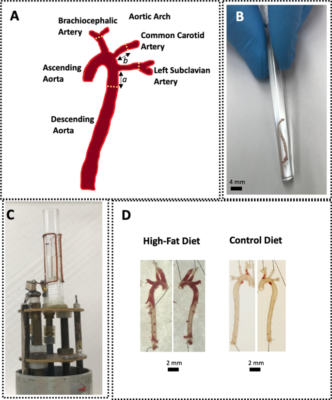
Materials and methods: Aortas from apolipoprotein-E-deficient (apoE-/-) mice fed an atherogenic diet (group 1) or a control diet (group 2) were subjected to 14 T MR imaging using a 3D gradient echo sequence. The obtained data sets were reconstructed (Matlab), segmented, and analyzed (Avizo). The aortas were further sectioned and subjected to traditional histological analysis (Oil-Red O and hematoxylin staining) for comparison.
Results: A resolution up to 15 × 10x10 μm3 revealed that plaque burden (mm3) was significantly (p < 0.05) higher in group 1 (0.41 ± 0.25, n = 4) than in group 2 (0.01 ± 0.01, n = 3). The achieved resolution provided similar detail on the plaque and the vessel wall morphology compared with histology. Digital image segmentation of the aorta's lumen, plaque, and wall offered three-dimensional visualizations of the entire, intact aortas.
Discussion: 14 T MR microscopy provided histology-like details of pathologically relevant vascular lesions. This work may provide the path research needs to take to enable plaque characterization in clinical applications.
期刊介绍:
MAGMA is a multidisciplinary international journal devoted to the publication of articles on all aspects of magnetic resonance techniques and their applications in medicine and biology. MAGMA currently publishes research papers, reviews, letters to the editor, and commentaries, six times a year. The subject areas covered by MAGMA include:
advances in materials, hardware and software in magnetic resonance technology,
new developments and results in research and practical applications of magnetic resonance imaging and spectroscopy related to biology and medicine,
study of animal models and intact cells using magnetic resonance,
reports of clinical trials on humans and clinical validation of magnetic resonance protocols.


 分享
分享
 求助内容:
求助内容: 应助结果提醒方式:
 扫码关注我们
