Layla Tahiri Elousrouti, Amal Mouaddine, Imane Fadlallah, Sofia Elhitmy, Sara Elloudi, Fatimazahra Mernissi, Mohammed Elidrissi, Nawal Hammas, Hinde Elfatemi, Laila Chbani
{"title":"原发性皮肤恶性血管周围细胞瘤Epithelioïd (PEComa): 1例报告并文献复习。","authors":"Layla Tahiri Elousrouti, Amal Mouaddine, Imane Fadlallah, Sofia Elhitmy, Sara Elloudi, Fatimazahra Mernissi, Mohammed Elidrissi, Nawal Hammas, Hinde Elfatemi, Laila Chbani","doi":"10.1177/2632010X231178629","DOIUrl":null,"url":null,"abstract":"<p><p>Perivascular epithelioïd cell tumor (PEComa) is a mesenchymal neoplasm with epithelioïd or spindled morphology with numerous thin-walled capillaries between tumor cells. They co-express markers of both melanocytic and smooth muscle differentiation. PEComas are rare, presenting in numerous anatomic sites including lung, kidney, liver, genitourinary tract, soft tissue, and skin. Primary cutaneous PEComas are very rare entity, and malignant ones are even more uncommon. Herein, we report the case of a 92-year-old female which was presenting with 7 cm exophytic, ulcerated, hemorrhagic nodular tumor, and rapidly growing for 8 months over the right thigh. On histologic examination, we found a dermal neoplasm formed by an atypical clear cell tumor with numerous branching capillaries between tumor cells. The mitotic count was found 6 mitotic figures/10 HPF. On immunohistochemistry, tumor cells co-expressed smooth muscle and melanocytic markers, CD10, and CD68. Based on these findings, the diagnosis of primary cutaneous malignant perivascular epithelioïd cell tumor (PEComa) was made. The large size (7 cm), the count of mitoses (6 mitotic figures/10 HPF), and the nuclear pleomorphism argued for malignancy. The absence of soft tissue or visceral localization argued for the cutaneous primitive origin. Adjuvant radiotherapy and targeted therapy with mTOR inhibitor (nab-sirolimus) was indicated. To the best of our knowledge, this is only the eighth case of a primary cutaneous malignant PEComa reported in the literature to date.</p>","PeriodicalId":72618,"journal":{"name":"","volume":"16 ","pages":"2632010X231178629"},"PeriodicalIF":0.0,"publicationDate":"2023-01-01","publicationTypes":"Journal Article","fieldsOfStudy":null,"isOpenAccess":false,"openAccessPdf":"https://ftp.ncbi.nlm.nih.gov/pub/pmc/oa_pdf/31/7d/10.1177_2632010X231178629.PMC10288419.pdf","citationCount":"0","resultStr":"{\"title\":\"Primary Cutaneous Malignant Perivascular Epithelioïd Cell Tumor (PEComa): Case Report With Review of the Literature.\",\"authors\":\"Layla Tahiri Elousrouti, Amal Mouaddine, Imane Fadlallah, Sofia Elhitmy, Sara Elloudi, Fatimazahra Mernissi, Mohammed Elidrissi, Nawal Hammas, Hinde Elfatemi, Laila Chbani\",\"doi\":\"10.1177/2632010X231178629\",\"DOIUrl\":null,\"url\":null,\"abstract\":\"<p><p>Perivascular epithelioïd cell tumor (PEComa) is a mesenchymal neoplasm with epithelioïd or spindled morphology with numerous thin-walled capillaries between tumor cells. They co-express markers of both melanocytic and smooth muscle differentiation. PEComas are rare, presenting in numerous anatomic sites including lung, kidney, liver, genitourinary tract, soft tissue, and skin. Primary cutaneous PEComas are very rare entity, and malignant ones are even more uncommon. Herein, we report the case of a 92-year-old female which was presenting with 7 cm exophytic, ulcerated, hemorrhagic nodular tumor, and rapidly growing for 8 months over the right thigh. On histologic examination, we found a dermal neoplasm formed by an atypical clear cell tumor with numerous branching capillaries between tumor cells. The mitotic count was found 6 mitotic figures/10 HPF. On immunohistochemistry, tumor cells co-expressed smooth muscle and melanocytic markers, CD10, and CD68. Based on these findings, the diagnosis of primary cutaneous malignant perivascular epithelioïd cell tumor (PEComa) was made. The large size (7 cm), the count of mitoses (6 mitotic figures/10 HPF), and the nuclear pleomorphism argued for malignancy. The absence of soft tissue or visceral localization argued for the cutaneous primitive origin. Adjuvant radiotherapy and targeted therapy with mTOR inhibitor (nab-sirolimus) was indicated. To the best of our knowledge, this is only the eighth case of a primary cutaneous malignant PEComa reported in the literature to date.</p>\",\"PeriodicalId\":72618,\"journal\":{\"name\":\"\",\"volume\":\"16 \",\"pages\":\"2632010X231178629\"},\"PeriodicalIF\":0.0,\"publicationDate\":\"2023-01-01\",\"publicationTypes\":\"Journal Article\",\"fieldsOfStudy\":null,\"isOpenAccess\":false,\"openAccessPdf\":\"https://ftp.ncbi.nlm.nih.gov/pub/pmc/oa_pdf/31/7d/10.1177_2632010X231178629.PMC10288419.pdf\",\"citationCount\":\"0\",\"resultStr\":null,\"platform\":\"Semanticscholar\",\"paperid\":null,\"PeriodicalName\":\"\",\"FirstCategoryId\":\"1085\",\"ListUrlMain\":\"https://doi.org/10.1177/2632010X231178629\",\"RegionNum\":0,\"RegionCategory\":null,\"ArticlePicture\":[],\"TitleCN\":null,\"AbstractTextCN\":null,\"PMCID\":null,\"EPubDate\":\"\",\"PubModel\":\"\",\"JCR\":\"\",\"JCRName\":\"\",\"Score\":null,\"Total\":0}","platform":"Semanticscholar","paperid":null,"PeriodicalName":"","FirstCategoryId":"1085","ListUrlMain":"https://doi.org/10.1177/2632010X231178629","RegionNum":0,"RegionCategory":null,"ArticlePicture":[],"TitleCN":null,"AbstractTextCN":null,"PMCID":null,"EPubDate":"","PubModel":"","JCR":"","JCRName":"","Score":null,"Total":0}
Primary Cutaneous Malignant Perivascular Epithelioïd Cell Tumor (PEComa): Case Report With Review of the Literature.
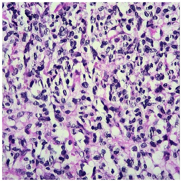
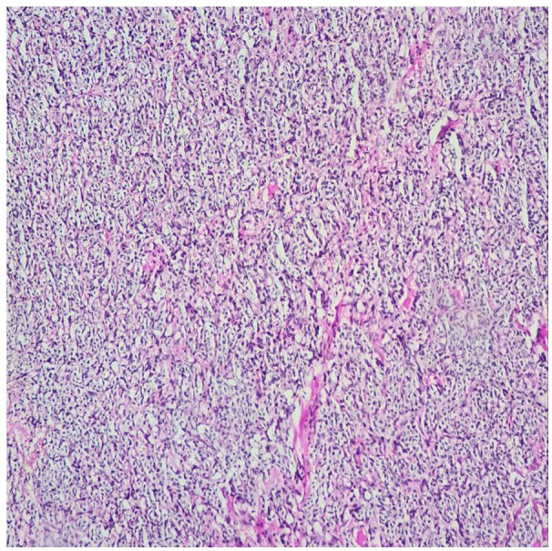
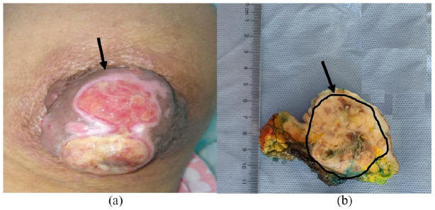
Perivascular epithelioïd cell tumor (PEComa) is a mesenchymal neoplasm with epithelioïd or spindled morphology with numerous thin-walled capillaries between tumor cells. They co-express markers of both melanocytic and smooth muscle differentiation. PEComas are rare, presenting in numerous anatomic sites including lung, kidney, liver, genitourinary tract, soft tissue, and skin. Primary cutaneous PEComas are very rare entity, and malignant ones are even more uncommon. Herein, we report the case of a 92-year-old female which was presenting with 7 cm exophytic, ulcerated, hemorrhagic nodular tumor, and rapidly growing for 8 months over the right thigh. On histologic examination, we found a dermal neoplasm formed by an atypical clear cell tumor with numerous branching capillaries between tumor cells. The mitotic count was found 6 mitotic figures/10 HPF. On immunohistochemistry, tumor cells co-expressed smooth muscle and melanocytic markers, CD10, and CD68. Based on these findings, the diagnosis of primary cutaneous malignant perivascular epithelioïd cell tumor (PEComa) was made. The large size (7 cm), the count of mitoses (6 mitotic figures/10 HPF), and the nuclear pleomorphism argued for malignancy. The absence of soft tissue or visceral localization argued for the cutaneous primitive origin. Adjuvant radiotherapy and targeted therapy with mTOR inhibitor (nab-sirolimus) was indicated. To the best of our knowledge, this is only the eighth case of a primary cutaneous malignant PEComa reported in the literature to date.


 分享
分享
 求助内容:
求助内容: 应助结果提醒方式:
 扫码关注我们
