Fahad H Alhazmi, Walaa M Alsharif, Sultan Abdulwadoud Alshoabi, Moawia Gameraddin, Khalid M Aloufi, Osama M Abdulaal, Abdualziz A Qurashi
{"title":"MRI识别COVID-19患者大脑微结构变化:一项系统综述","authors":"Fahad H Alhazmi, Walaa M Alsharif, Sultan Abdulwadoud Alshoabi, Moawia Gameraddin, Khalid M Aloufi, Osama M Abdulaal, Abdualziz A Qurashi","doi":"10.4103/bc.bc_77_22","DOIUrl":null,"url":null,"abstract":"<p><p>Coronavirus disease 2019 (COVID-19) is an epidemic viral disease caused by a novel severe acute respiratory syndrome coronavirus 2 (SARS-CoV-2). Despite the excessive number of neurological articles that have investigated the effect of COVID-19 on the brain from the neurological point of view, very few studies have investigated the impact of COVID-19 on the cerebral microstructure and function of the brain. The aim of this study was to summarize the results of the existing studies on cerebral microstructural changes in COVID-19 patients, specifically the use of quantitative volumetric analysis, blood oxygen level dependent (BOLD), and diffusion tensor imaging (DTI). We searched PubMed/MEDLINE, ScienceDirect, Semantic Scholar, and Google Scholar from December 2020 to April 2022. A well-constructed search strategy was used to identify the articles for review. Seven research articles have met this study's inclusion and exclusion criteria, which have applied neuroimaging tools such as quantitative volumetric analysis, BOLD, and DTI to investigate cerebral microstructure changes in COVID-19 patients. A significant effect of COVID-19 was found in the brain such as hypoperfusion of cerebral blood flow, increased gray matter (GM) volume, and reduced cortical thickness. The insula and thalamic radiation were the most frequent GM region and white matter tract, respectively, that are involved in SARS-CoV-2. COVID-19 was found to be associated with changes in cerebral microstructures. These abnormalities in brain areas might lead to be associated with behaviors, mental and neurological alterations that need to be considered carefully in future studies.</p>","PeriodicalId":9288,"journal":{"name":"Brain Circulation","volume":"9 1","pages":"6-15"},"PeriodicalIF":4.8000,"publicationDate":"2023-01-01","publicationTypes":"Journal Article","fieldsOfStudy":null,"isOpenAccess":false,"openAccessPdf":"https://ftp.ncbi.nlm.nih.gov/pub/pmc/oa_pdf/20/02/BC-9-6.PMC10158661.pdf","citationCount":"0","resultStr":"{\"title\":\"Identifying cerebral microstructural changes in patients with COVID-19 using MRI: A systematic review.\",\"authors\":\"Fahad H Alhazmi, Walaa M Alsharif, Sultan Abdulwadoud Alshoabi, Moawia Gameraddin, Khalid M Aloufi, Osama M Abdulaal, Abdualziz A Qurashi\",\"doi\":\"10.4103/bc.bc_77_22\",\"DOIUrl\":null,\"url\":null,\"abstract\":\"<p><p>Coronavirus disease 2019 (COVID-19) is an epidemic viral disease caused by a novel severe acute respiratory syndrome coronavirus 2 (SARS-CoV-2). Despite the excessive number of neurological articles that have investigated the effect of COVID-19 on the brain from the neurological point of view, very few studies have investigated the impact of COVID-19 on the cerebral microstructure and function of the brain. The aim of this study was to summarize the results of the existing studies on cerebral microstructural changes in COVID-19 patients, specifically the use of quantitative volumetric analysis, blood oxygen level dependent (BOLD), and diffusion tensor imaging (DTI). We searched PubMed/MEDLINE, ScienceDirect, Semantic Scholar, and Google Scholar from December 2020 to April 2022. A well-constructed search strategy was used to identify the articles for review. Seven research articles have met this study's inclusion and exclusion criteria, which have applied neuroimaging tools such as quantitative volumetric analysis, BOLD, and DTI to investigate cerebral microstructure changes in COVID-19 patients. A significant effect of COVID-19 was found in the brain such as hypoperfusion of cerebral blood flow, increased gray matter (GM) volume, and reduced cortical thickness. The insula and thalamic radiation were the most frequent GM region and white matter tract, respectively, that are involved in SARS-CoV-2. COVID-19 was found to be associated with changes in cerebral microstructures. These abnormalities in brain areas might lead to be associated with behaviors, mental and neurological alterations that need to be considered carefully in future studies.</p>\",\"PeriodicalId\":9288,\"journal\":{\"name\":\"Brain Circulation\",\"volume\":\"9 1\",\"pages\":\"6-15\"},\"PeriodicalIF\":4.8000,\"publicationDate\":\"2023-01-01\",\"publicationTypes\":\"Journal Article\",\"fieldsOfStudy\":null,\"isOpenAccess\":false,\"openAccessPdf\":\"https://ftp.ncbi.nlm.nih.gov/pub/pmc/oa_pdf/20/02/BC-9-6.PMC10158661.pdf\",\"citationCount\":\"0\",\"resultStr\":null,\"platform\":\"Semanticscholar\",\"paperid\":null,\"PeriodicalName\":\"Brain Circulation\",\"FirstCategoryId\":\"3\",\"ListUrlMain\":\"https://doi.org/10.4103/bc.bc_77_22\",\"RegionNum\":4,\"RegionCategory\":\"医学\",\"ArticlePicture\":[],\"TitleCN\":null,\"AbstractTextCN\":null,\"PMCID\":null,\"EPubDate\":\"\",\"PubModel\":\"\",\"JCR\":\"Q3\",\"JCRName\":\"CLINICAL NEUROLOGY\",\"Score\":null,\"Total\":0}","platform":"Semanticscholar","paperid":null,"PeriodicalName":"Brain Circulation","FirstCategoryId":"3","ListUrlMain":"https://doi.org/10.4103/bc.bc_77_22","RegionNum":4,"RegionCategory":"医学","ArticlePicture":[],"TitleCN":null,"AbstractTextCN":null,"PMCID":null,"EPubDate":"","PubModel":"","JCR":"Q3","JCRName":"CLINICAL NEUROLOGY","Score":null,"Total":0}
Identifying cerebral microstructural changes in patients with COVID-19 using MRI: A systematic review.
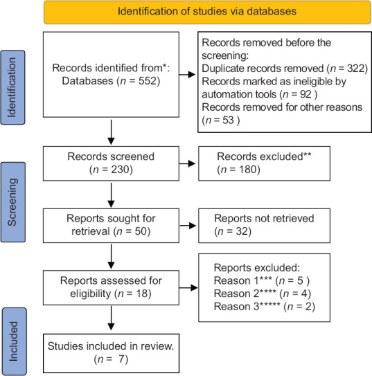
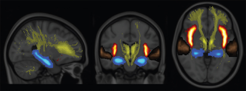
Coronavirus disease 2019 (COVID-19) is an epidemic viral disease caused by a novel severe acute respiratory syndrome coronavirus 2 (SARS-CoV-2). Despite the excessive number of neurological articles that have investigated the effect of COVID-19 on the brain from the neurological point of view, very few studies have investigated the impact of COVID-19 on the cerebral microstructure and function of the brain. The aim of this study was to summarize the results of the existing studies on cerebral microstructural changes in COVID-19 patients, specifically the use of quantitative volumetric analysis, blood oxygen level dependent (BOLD), and diffusion tensor imaging (DTI). We searched PubMed/MEDLINE, ScienceDirect, Semantic Scholar, and Google Scholar from December 2020 to April 2022. A well-constructed search strategy was used to identify the articles for review. Seven research articles have met this study's inclusion and exclusion criteria, which have applied neuroimaging tools such as quantitative volumetric analysis, BOLD, and DTI to investigate cerebral microstructure changes in COVID-19 patients. A significant effect of COVID-19 was found in the brain such as hypoperfusion of cerebral blood flow, increased gray matter (GM) volume, and reduced cortical thickness. The insula and thalamic radiation were the most frequent GM region and white matter tract, respectively, that are involved in SARS-CoV-2. COVID-19 was found to be associated with changes in cerebral microstructures. These abnormalities in brain areas might lead to be associated with behaviors, mental and neurological alterations that need to be considered carefully in future studies.


 分享
分享
 求助内容:
求助内容: 应助结果提醒方式:
 扫码关注我们
