{"title":"心脏计算机断层扫描血管造影术检测主动脉瓣叶动脉瘤的独特病例。","authors":"Valeria Pergola, Giulio Cabrelle, Raffaella Motta","doi":"10.4103/jcecho.jcecho_59_22","DOIUrl":null,"url":null,"abstract":"<p><p>Heart valve leaflet's aneurysm is a rare finding, and literature about this topic is sparse. Early recognition is important because their rupture can lead to catastrophic valve regurgitation. An 84-year-old male with chronic ischemic cardiomyopathy was admitted to the coronary intensive care unit for non-ST elevation myocardial infarction. Baseline transthoracic echocardiography showed normal biventricular function with inhomogeneous thickening of aortic leaflets with moderate aortic regurgitation. Because the acoustic window was limited, a transesophageal echocardiography was performed, detecting a small mass in the right aortic coronary cusp with moderate regurgitation (orifice regurgitation area: 0.54 cm<sup>2</sup>; med/max gradient: 16/32 mmHg). Endocarditis was ruled out. Because of the rapid worsening of the patient's conditions, requiring mechanical ventilation and hemofiltration, and the potential hazard of an urgent coronary angiography, a cardiac computed tomographic angiography was performed. Detailed spatial reconstructions highlighted a bilobed cavitation in the aortic leaflets. Diagnosis of aortic leaflets' aneurysm was made. A \"wait and see\" strategy was chosen, and the patient's general conditions gradually improved and now he is stable and uneventful. To date, no aortic leaflet's aneurysm was described in literature.</p>","PeriodicalId":15191,"journal":{"name":"Journal of Cardiovascular Echography","volume":"33 1","pages":"30-32"},"PeriodicalIF":1.0000,"publicationDate":"2023-01-01","publicationTypes":"Journal Article","fieldsOfStudy":null,"isOpenAccess":false,"openAccessPdf":"https://www.ncbi.nlm.nih.gov/pmc/articles/PMC10328130/pdf/","citationCount":"0","resultStr":"{\"title\":\"A Unique Case of Aortic Valve Leaflet's Aneurysm Detected by Cardiac Computed Tomographic Angiography.\",\"authors\":\"Valeria Pergola, Giulio Cabrelle, Raffaella Motta\",\"doi\":\"10.4103/jcecho.jcecho_59_22\",\"DOIUrl\":null,\"url\":null,\"abstract\":\"<p><p>Heart valve leaflet's aneurysm is a rare finding, and literature about this topic is sparse. Early recognition is important because their rupture can lead to catastrophic valve regurgitation. An 84-year-old male with chronic ischemic cardiomyopathy was admitted to the coronary intensive care unit for non-ST elevation myocardial infarction. Baseline transthoracic echocardiography showed normal biventricular function with inhomogeneous thickening of aortic leaflets with moderate aortic regurgitation. Because the acoustic window was limited, a transesophageal echocardiography was performed, detecting a small mass in the right aortic coronary cusp with moderate regurgitation (orifice regurgitation area: 0.54 cm<sup>2</sup>; med/max gradient: 16/32 mmHg). Endocarditis was ruled out. Because of the rapid worsening of the patient's conditions, requiring mechanical ventilation and hemofiltration, and the potential hazard of an urgent coronary angiography, a cardiac computed tomographic angiography was performed. Detailed spatial reconstructions highlighted a bilobed cavitation in the aortic leaflets. Diagnosis of aortic leaflets' aneurysm was made. A \\\"wait and see\\\" strategy was chosen, and the patient's general conditions gradually improved and now he is stable and uneventful. To date, no aortic leaflet's aneurysm was described in literature.</p>\",\"PeriodicalId\":15191,\"journal\":{\"name\":\"Journal of Cardiovascular Echography\",\"volume\":\"33 1\",\"pages\":\"30-32\"},\"PeriodicalIF\":1.0000,\"publicationDate\":\"2023-01-01\",\"publicationTypes\":\"Journal Article\",\"fieldsOfStudy\":null,\"isOpenAccess\":false,\"openAccessPdf\":\"https://www.ncbi.nlm.nih.gov/pmc/articles/PMC10328130/pdf/\",\"citationCount\":\"0\",\"resultStr\":null,\"platform\":\"Semanticscholar\",\"paperid\":null,\"PeriodicalName\":\"Journal of Cardiovascular Echography\",\"FirstCategoryId\":\"1085\",\"ListUrlMain\":\"https://doi.org/10.4103/jcecho.jcecho_59_22\",\"RegionNum\":0,\"RegionCategory\":null,\"ArticlePicture\":[],\"TitleCN\":null,\"AbstractTextCN\":null,\"PMCID\":null,\"EPubDate\":\"2023/5/29 0:00:00\",\"PubModel\":\"Epub\",\"JCR\":\"Q4\",\"JCRName\":\"CARDIAC & CARDIOVASCULAR SYSTEMS\",\"Score\":null,\"Total\":0}","platform":"Semanticscholar","paperid":null,"PeriodicalName":"Journal of Cardiovascular Echography","FirstCategoryId":"1085","ListUrlMain":"https://doi.org/10.4103/jcecho.jcecho_59_22","RegionNum":0,"RegionCategory":null,"ArticlePicture":[],"TitleCN":null,"AbstractTextCN":null,"PMCID":null,"EPubDate":"2023/5/29 0:00:00","PubModel":"Epub","JCR":"Q4","JCRName":"CARDIAC & CARDIOVASCULAR SYSTEMS","Score":null,"Total":0}
A Unique Case of Aortic Valve Leaflet's Aneurysm Detected by Cardiac Computed Tomographic Angiography.
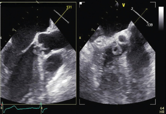
Heart valve leaflet's aneurysm is a rare finding, and literature about this topic is sparse. Early recognition is important because their rupture can lead to catastrophic valve regurgitation. An 84-year-old male with chronic ischemic cardiomyopathy was admitted to the coronary intensive care unit for non-ST elevation myocardial infarction. Baseline transthoracic echocardiography showed normal biventricular function with inhomogeneous thickening of aortic leaflets with moderate aortic regurgitation. Because the acoustic window was limited, a transesophageal echocardiography was performed, detecting a small mass in the right aortic coronary cusp with moderate regurgitation (orifice regurgitation area: 0.54 cm2; med/max gradient: 16/32 mmHg). Endocarditis was ruled out. Because of the rapid worsening of the patient's conditions, requiring mechanical ventilation and hemofiltration, and the potential hazard of an urgent coronary angiography, a cardiac computed tomographic angiography was performed. Detailed spatial reconstructions highlighted a bilobed cavitation in the aortic leaflets. Diagnosis of aortic leaflets' aneurysm was made. A "wait and see" strategy was chosen, and the patient's general conditions gradually improved and now he is stable and uneventful. To date, no aortic leaflet's aneurysm was described in literature.


 分享
分享
 求助内容:
求助内容: 应助结果提醒方式:
 扫码关注我们
