Şevket Kahraman, Mesut Furkan Yazar, Hüseyin Aydemir, Mecit Kantarci, Sonay Aydin
{"title":"计算机断层扫描检测气管分支:角度与年龄性别的关系。","authors":"Şevket Kahraman, Mesut Furkan Yazar, Hüseyin Aydemir, Mecit Kantarci, Sonay Aydin","doi":"10.4329/wjr.v15.i4.118","DOIUrl":null,"url":null,"abstract":"<p><strong>Background: </strong>The data obtained on the anatomical knowledge of the tracheobronchial system can be used for diagnosis, treatment and interventional interventions in areas such as anesthesia, thoracic surgery, pulmonary physiology.</p><p><strong>Aim: </strong>To determine the tracheobronchial branching angles in pediatric and adult populations by using the multislice computed tomography (CT) and minimum intensity projection (MinIP) technique, which is a non-invasive method.</p><p><strong>Methods: </strong>Our study was carried out retrospectively. Patients who underwent contrast and non-contrast CT examination, whose anatomically and pathophysiologically good tracheobronchial system and lung parenchyma images were obtained, were included in the study. Measurements were made in the coronal plane of the lung parenchyma. In the coronal plane, right main bronchus-left main bronchus angle, right upper lobe bronchus-intermedius bronchus angle, right middle lobe bronchus-right lower lobe bronchus angle, left upper lobe bronchus-left lower lobe bronchus angle were measured.</p><p><strong>Results: </strong>The study population consisted of 1511 patients, 753 pediatric (mean age: 13.4 ± 4.3; range: 1-18 years) and 758 adults (mean age: 54.3 ± 17.3; range: 19-94 years). In our study, tracheal bifurcation angle was found to be 73.3° ± 13.7° (59.6°-87°) in the whole population. In the pediatric group, the right-left main coronal level was found to be higher in boys compared to girls (74.6° ± 12.9° <i>vs</i> 71.2° ± 13.9°, <i>P</i> = 0.001). In the adult group, the right-left main coronal level was found to be lower in males compared to females (71.9° ± 12.9° <i>vs</i> 75.8° ± 14.7°, <i>P</i> < 0.001).</p><p><strong>Conclusions: </strong>Our study, with the number of 1511 patients, is the first study in the literature with the largest number of patient populations including pediatric and adult demographic data, measuring the angle values of the tracheobronchial system using multislice CT and MinIP technique. Study data will not only be a guide during invasive procedures, but it can also guide studies to be done with imaging methods.</p>","PeriodicalId":23819,"journal":{"name":"World journal of radiology","volume":"15 4","pages":"118-126"},"PeriodicalIF":1.5000,"publicationDate":"2023-04-28","publicationTypes":"Journal Article","fieldsOfStudy":null,"isOpenAccess":false,"openAccessPdf":"https://ftp.ncbi.nlm.nih.gov/pub/pmc/oa_pdf/a1/b3/WJR-15-118.PMC10167816.pdf","citationCount":"0","resultStr":"{\"title\":\"Detection of tracheal branching with computerized tomography: The relationship between the angles and age-gender.\",\"authors\":\"Şevket Kahraman, Mesut Furkan Yazar, Hüseyin Aydemir, Mecit Kantarci, Sonay Aydin\",\"doi\":\"10.4329/wjr.v15.i4.118\",\"DOIUrl\":null,\"url\":null,\"abstract\":\"<p><strong>Background: </strong>The data obtained on the anatomical knowledge of the tracheobronchial system can be used for diagnosis, treatment and interventional interventions in areas such as anesthesia, thoracic surgery, pulmonary physiology.</p><p><strong>Aim: </strong>To determine the tracheobronchial branching angles in pediatric and adult populations by using the multislice computed tomography (CT) and minimum intensity projection (MinIP) technique, which is a non-invasive method.</p><p><strong>Methods: </strong>Our study was carried out retrospectively. Patients who underwent contrast and non-contrast CT examination, whose anatomically and pathophysiologically good tracheobronchial system and lung parenchyma images were obtained, were included in the study. Measurements were made in the coronal plane of the lung parenchyma. In the coronal plane, right main bronchus-left main bronchus angle, right upper lobe bronchus-intermedius bronchus angle, right middle lobe bronchus-right lower lobe bronchus angle, left upper lobe bronchus-left lower lobe bronchus angle were measured.</p><p><strong>Results: </strong>The study population consisted of 1511 patients, 753 pediatric (mean age: 13.4 ± 4.3; range: 1-18 years) and 758 adults (mean age: 54.3 ± 17.3; range: 19-94 years). In our study, tracheal bifurcation angle was found to be 73.3° ± 13.7° (59.6°-87°) in the whole population. In the pediatric group, the right-left main coronal level was found to be higher in boys compared to girls (74.6° ± 12.9° <i>vs</i> 71.2° ± 13.9°, <i>P</i> = 0.001). In the adult group, the right-left main coronal level was found to be lower in males compared to females (71.9° ± 12.9° <i>vs</i> 75.8° ± 14.7°, <i>P</i> < 0.001).</p><p><strong>Conclusions: </strong>Our study, with the number of 1511 patients, is the first study in the literature with the largest number of patient populations including pediatric and adult demographic data, measuring the angle values of the tracheobronchial system using multislice CT and MinIP technique. Study data will not only be a guide during invasive procedures, but it can also guide studies to be done with imaging methods.</p>\",\"PeriodicalId\":23819,\"journal\":{\"name\":\"World journal of radiology\",\"volume\":\"15 4\",\"pages\":\"118-126\"},\"PeriodicalIF\":1.5000,\"publicationDate\":\"2023-04-28\",\"publicationTypes\":\"Journal Article\",\"fieldsOfStudy\":null,\"isOpenAccess\":false,\"openAccessPdf\":\"https://ftp.ncbi.nlm.nih.gov/pub/pmc/oa_pdf/a1/b3/WJR-15-118.PMC10167816.pdf\",\"citationCount\":\"0\",\"resultStr\":null,\"platform\":\"Semanticscholar\",\"paperid\":null,\"PeriodicalName\":\"World journal of radiology\",\"FirstCategoryId\":\"1085\",\"ListUrlMain\":\"https://doi.org/10.4329/wjr.v15.i4.118\",\"RegionNum\":0,\"RegionCategory\":null,\"ArticlePicture\":[],\"TitleCN\":null,\"AbstractTextCN\":null,\"PMCID\":null,\"EPubDate\":\"\",\"PubModel\":\"\",\"JCR\":\"Q3\",\"JCRName\":\"RADIOLOGY, NUCLEAR MEDICINE & MEDICAL IMAGING\",\"Score\":null,\"Total\":0}","platform":"Semanticscholar","paperid":null,"PeriodicalName":"World journal of radiology","FirstCategoryId":"1085","ListUrlMain":"https://doi.org/10.4329/wjr.v15.i4.118","RegionNum":0,"RegionCategory":null,"ArticlePicture":[],"TitleCN":null,"AbstractTextCN":null,"PMCID":null,"EPubDate":"","PubModel":"","JCR":"Q3","JCRName":"RADIOLOGY, NUCLEAR MEDICINE & MEDICAL IMAGING","Score":null,"Total":0}
Detection of tracheal branching with computerized tomography: The relationship between the angles and age-gender.
Background: The data obtained on the anatomical knowledge of the tracheobronchial system can be used for diagnosis, treatment and interventional interventions in areas such as anesthesia, thoracic surgery, pulmonary physiology.
Aim: To determine the tracheobronchial branching angles in pediatric and adult populations by using the multislice computed tomography (CT) and minimum intensity projection (MinIP) technique, which is a non-invasive method.
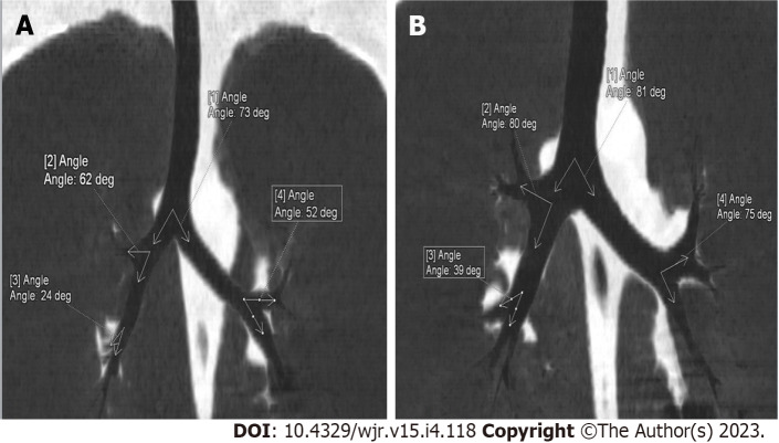
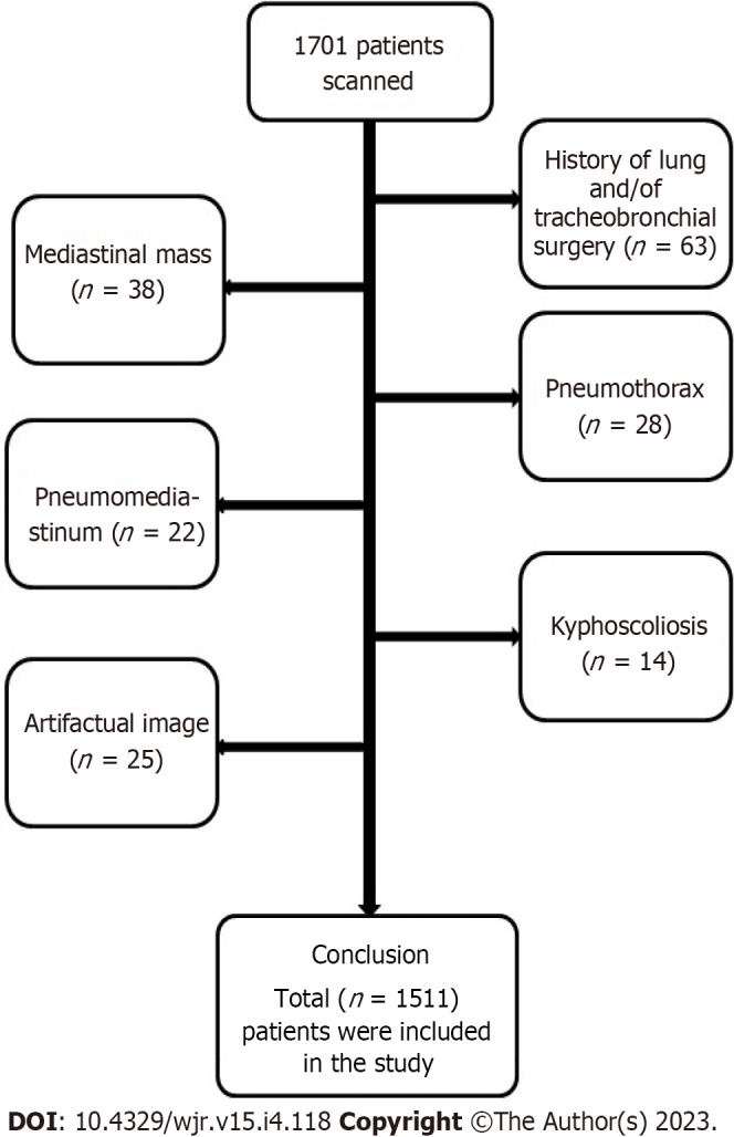
Methods: Our study was carried out retrospectively. Patients who underwent contrast and non-contrast CT examination, whose anatomically and pathophysiologically good tracheobronchial system and lung parenchyma images were obtained, were included in the study. Measurements were made in the coronal plane of the lung parenchyma. In the coronal plane, right main bronchus-left main bronchus angle, right upper lobe bronchus-intermedius bronchus angle, right middle lobe bronchus-right lower lobe bronchus angle, left upper lobe bronchus-left lower lobe bronchus angle were measured.
Results: The study population consisted of 1511 patients, 753 pediatric (mean age: 13.4 ± 4.3; range: 1-18 years) and 758 adults (mean age: 54.3 ± 17.3; range: 19-94 years). In our study, tracheal bifurcation angle was found to be 73.3° ± 13.7° (59.6°-87°) in the whole population. In the pediatric group, the right-left main coronal level was found to be higher in boys compared to girls (74.6° ± 12.9° vs 71.2° ± 13.9°, P = 0.001). In the adult group, the right-left main coronal level was found to be lower in males compared to females (71.9° ± 12.9° vs 75.8° ± 14.7°, P < 0.001).
Conclusions: Our study, with the number of 1511 patients, is the first study in the literature with the largest number of patient populations including pediatric and adult demographic data, measuring the angle values of the tracheobronchial system using multislice CT and MinIP technique. Study data will not only be a guide during invasive procedures, but it can also guide studies to be done with imaging methods.


 分享
分享
 求助内容:
求助内容: 应助结果提醒方式:
 扫码关注我们
