{"title":"急性大脑中动脉闭塞反复经皮腔内血管成形术后造影剂所致脑病1例。","authors":"Haruki Otsubo, Tomohide Yoshie, Takashi Araga, Kentaro Tatsuno, Satoshi Takaishi, Noriko Usuki, Yasuyuki Yoshida, Hajime Ono, Toshihiro Ueda","doi":"10.5797/jnet.cr.2021-0061","DOIUrl":null,"url":null,"abstract":"<p><strong>Objective: </strong>We report a case of contrast-induced encephalopathy (CIE) after repeated percutaneous transluminal angioplasty (PTA) for acute middle cerebral artery (MCA) occlusion.</p><p><strong>Case presentation: </strong>An 88-year-old woman with left hemiparesis was transferred to our hospital by ambulance. MRI revealed acute MCA M1 occlusion. We performed intravenous tissue plasminogen activator therapy and PTA for right MCA occlusion, leading to complete recanalization and improvement in hemiparalysis. After approximately one week, restenosis of right MCA developed and PTA was performed again on day 11. However, her left hemiparesis exacerbated shortly thereafter. CT demonstrated leakage of contrast medium, and an extensive high-intensity area (HIA) on the white matter in the right cerebral hemisphere was noted on MRI FLAIR. The HIA on MRI and neurological deficits gradually improved after conservative treatment, but diffuse atrophy of the right cerebral hemisphere occurred and higher brain dysfunction remained.</p><p><strong>Conclusion: </strong>Repeated ischemia and reperfusion, and the frequent use of contrast media were considered the causes of CIE.</p>","PeriodicalId":73856,"journal":{"name":"Journal of neuroendovascular therapy","volume":"16 7","pages":"371-375"},"PeriodicalIF":0.5000,"publicationDate":"2022-01-01","publicationTypes":"Journal Article","fieldsOfStudy":null,"isOpenAccess":false,"openAccessPdf":"https://ftp.ncbi.nlm.nih.gov/pub/pmc/oa_pdf/36/7b/jnet-16-371.PMC10370915.pdf","citationCount":"0","resultStr":"{\"title\":\"A Case Report of Contrast-Induced Encephalopathy after Repeated Percutaneous Transluminal Angioplasty for Acute Middle Cerebral Artery Occlusion.\",\"authors\":\"Haruki Otsubo, Tomohide Yoshie, Takashi Araga, Kentaro Tatsuno, Satoshi Takaishi, Noriko Usuki, Yasuyuki Yoshida, Hajime Ono, Toshihiro Ueda\",\"doi\":\"10.5797/jnet.cr.2021-0061\",\"DOIUrl\":null,\"url\":null,\"abstract\":\"<p><strong>Objective: </strong>We report a case of contrast-induced encephalopathy (CIE) after repeated percutaneous transluminal angioplasty (PTA) for acute middle cerebral artery (MCA) occlusion.</p><p><strong>Case presentation: </strong>An 88-year-old woman with left hemiparesis was transferred to our hospital by ambulance. MRI revealed acute MCA M1 occlusion. We performed intravenous tissue plasminogen activator therapy and PTA for right MCA occlusion, leading to complete recanalization and improvement in hemiparalysis. After approximately one week, restenosis of right MCA developed and PTA was performed again on day 11. However, her left hemiparesis exacerbated shortly thereafter. CT demonstrated leakage of contrast medium, and an extensive high-intensity area (HIA) on the white matter in the right cerebral hemisphere was noted on MRI FLAIR. The HIA on MRI and neurological deficits gradually improved after conservative treatment, but diffuse atrophy of the right cerebral hemisphere occurred and higher brain dysfunction remained.</p><p><strong>Conclusion: </strong>Repeated ischemia and reperfusion, and the frequent use of contrast media were considered the causes of CIE.</p>\",\"PeriodicalId\":73856,\"journal\":{\"name\":\"Journal of neuroendovascular therapy\",\"volume\":\"16 7\",\"pages\":\"371-375\"},\"PeriodicalIF\":0.5000,\"publicationDate\":\"2022-01-01\",\"publicationTypes\":\"Journal Article\",\"fieldsOfStudy\":null,\"isOpenAccess\":false,\"openAccessPdf\":\"https://ftp.ncbi.nlm.nih.gov/pub/pmc/oa_pdf/36/7b/jnet-16-371.PMC10370915.pdf\",\"citationCount\":\"0\",\"resultStr\":null,\"platform\":\"Semanticscholar\",\"paperid\":null,\"PeriodicalName\":\"Journal of neuroendovascular therapy\",\"FirstCategoryId\":\"1085\",\"ListUrlMain\":\"https://doi.org/10.5797/jnet.cr.2021-0061\",\"RegionNum\":0,\"RegionCategory\":null,\"ArticlePicture\":[],\"TitleCN\":null,\"AbstractTextCN\":null,\"PMCID\":null,\"EPubDate\":\"\",\"PubModel\":\"\",\"JCR\":\"\",\"JCRName\":\"\",\"Score\":null,\"Total\":0}","platform":"Semanticscholar","paperid":null,"PeriodicalName":"Journal of neuroendovascular therapy","FirstCategoryId":"1085","ListUrlMain":"https://doi.org/10.5797/jnet.cr.2021-0061","RegionNum":0,"RegionCategory":null,"ArticlePicture":[],"TitleCN":null,"AbstractTextCN":null,"PMCID":null,"EPubDate":"","PubModel":"","JCR":"","JCRName":"","Score":null,"Total":0}
A Case Report of Contrast-Induced Encephalopathy after Repeated Percutaneous Transluminal Angioplasty for Acute Middle Cerebral Artery Occlusion.
Objective: We report a case of contrast-induced encephalopathy (CIE) after repeated percutaneous transluminal angioplasty (PTA) for acute middle cerebral artery (MCA) occlusion.
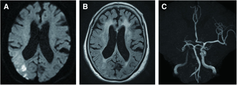
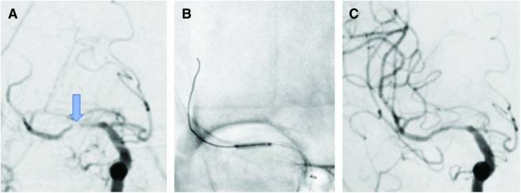
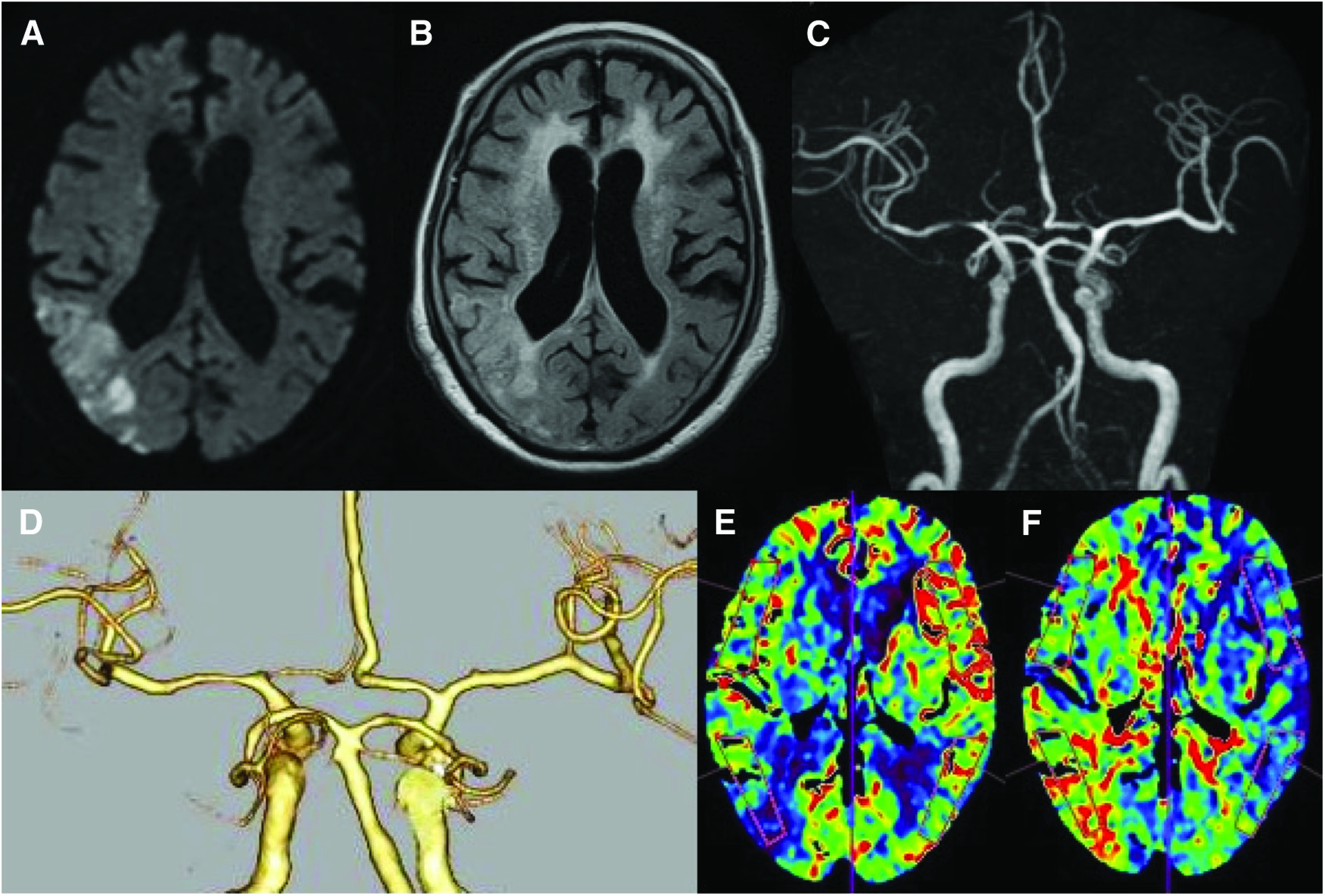
Case presentation: An 88-year-old woman with left hemiparesis was transferred to our hospital by ambulance. MRI revealed acute MCA M1 occlusion. We performed intravenous tissue plasminogen activator therapy and PTA for right MCA occlusion, leading to complete recanalization and improvement in hemiparalysis. After approximately one week, restenosis of right MCA developed and PTA was performed again on day 11. However, her left hemiparesis exacerbated shortly thereafter. CT demonstrated leakage of contrast medium, and an extensive high-intensity area (HIA) on the white matter in the right cerebral hemisphere was noted on MRI FLAIR. The HIA on MRI and neurological deficits gradually improved after conservative treatment, but diffuse atrophy of the right cerebral hemisphere occurred and higher brain dysfunction remained.
Conclusion: Repeated ischemia and reperfusion, and the frequent use of contrast media were considered the causes of CIE.


 分享
分享

 求助内容:
求助内容: 应助结果提醒方式:
 扫码关注我们
