{"title":"伴有玻璃体种子的恶性脉络膜黑色素瘤:由组织病理学和场发射扫描电子显微镜研究支持。","authors":"Dipankar Das, Obaidur Rehman, Kasturi Bhattacharjee, Harsha Bhattacharjee, Manab Jyoti Barman, Surjendu Maity, Dipankar Bandyopadhyay","doi":"10.22336/rjo.2023.31","DOIUrl":null,"url":null,"abstract":"<p><p><b>Aim:</b> To report an exceptionally rare case of malignant choroidal melanoma with vitreous seeding, supported by histopathological and field emission scanning electron microscopic (FESEM) studies. <b>Case report:</b> A 58-year-old male with painless diminution of vision in his left eye for past 1 month was found to have a brown retrolental mass lesion on slit lamp examination in the left eye. Detailed fundus examination revealed choroidal melanoma in the left eye with pigmented seeds extending into the vitreous cavity and associated exudative retinal detachment. Ocular imaging was consistent with the diagnosis. <b>Results:</b> The eyeball was enucleated and the tumor was considered as stage IIB (AJCC 8th edition classification). Metastatic workup of the patient was negative. One half of the eyeball was subjected to field emission scanning electron microscopy to further study the nature and appearance of vitreous seeds. <b>Discussion:</b> Vitreous seeding in choroidal melanoma has been reported only in a handful of cases in literature. Histopathological confirmation of vitreous seeds was done in our case and morphological detailing was performed using FESEM study. <b>Conclusions:</b> Treatment naïve choroidal melanoma can very rarely have vitreous seeds. Early enucleation in such cases carries a favorable prognosis.</p>","PeriodicalId":21385,"journal":{"name":"Romanian journal of ophthalmology","volume":"67 2","pages":"180-184"},"PeriodicalIF":0.0000,"publicationDate":"2023-04-01","publicationTypes":"Journal Article","fieldsOfStudy":null,"isOpenAccess":false,"openAccessPdf":"https://www.ncbi.nlm.nih.gov/pmc/articles/PMC10385716/pdf/","citationCount":"0","resultStr":"{\"title\":\"Malignant choroidal melanoma with vitreous seeds: supported by histopathology and field emission scanning electron microscopy study.\",\"authors\":\"Dipankar Das, Obaidur Rehman, Kasturi Bhattacharjee, Harsha Bhattacharjee, Manab Jyoti Barman, Surjendu Maity, Dipankar Bandyopadhyay\",\"doi\":\"10.22336/rjo.2023.31\",\"DOIUrl\":null,\"url\":null,\"abstract\":\"<p><p><b>Aim:</b> To report an exceptionally rare case of malignant choroidal melanoma with vitreous seeding, supported by histopathological and field emission scanning electron microscopic (FESEM) studies. <b>Case report:</b> A 58-year-old male with painless diminution of vision in his left eye for past 1 month was found to have a brown retrolental mass lesion on slit lamp examination in the left eye. Detailed fundus examination revealed choroidal melanoma in the left eye with pigmented seeds extending into the vitreous cavity and associated exudative retinal detachment. Ocular imaging was consistent with the diagnosis. <b>Results:</b> The eyeball was enucleated and the tumor was considered as stage IIB (AJCC 8th edition classification). Metastatic workup of the patient was negative. One half of the eyeball was subjected to field emission scanning electron microscopy to further study the nature and appearance of vitreous seeds. <b>Discussion:</b> Vitreous seeding in choroidal melanoma has been reported only in a handful of cases in literature. Histopathological confirmation of vitreous seeds was done in our case and morphological detailing was performed using FESEM study. <b>Conclusions:</b> Treatment naïve choroidal melanoma can very rarely have vitreous seeds. Early enucleation in such cases carries a favorable prognosis.</p>\",\"PeriodicalId\":21385,\"journal\":{\"name\":\"Romanian journal of ophthalmology\",\"volume\":\"67 2\",\"pages\":\"180-184\"},\"PeriodicalIF\":0.0000,\"publicationDate\":\"2023-04-01\",\"publicationTypes\":\"Journal Article\",\"fieldsOfStudy\":null,\"isOpenAccess\":false,\"openAccessPdf\":\"https://www.ncbi.nlm.nih.gov/pmc/articles/PMC10385716/pdf/\",\"citationCount\":\"0\",\"resultStr\":null,\"platform\":\"Semanticscholar\",\"paperid\":null,\"PeriodicalName\":\"Romanian journal of ophthalmology\",\"FirstCategoryId\":\"1085\",\"ListUrlMain\":\"https://doi.org/10.22336/rjo.2023.31\",\"RegionNum\":0,\"RegionCategory\":null,\"ArticlePicture\":[],\"TitleCN\":null,\"AbstractTextCN\":null,\"PMCID\":null,\"EPubDate\":\"\",\"PubModel\":\"\",\"JCR\":\"\",\"JCRName\":\"\",\"Score\":null,\"Total\":0}","platform":"Semanticscholar","paperid":null,"PeriodicalName":"Romanian journal of ophthalmology","FirstCategoryId":"1085","ListUrlMain":"https://doi.org/10.22336/rjo.2023.31","RegionNum":0,"RegionCategory":null,"ArticlePicture":[],"TitleCN":null,"AbstractTextCN":null,"PMCID":null,"EPubDate":"","PubModel":"","JCR":"","JCRName":"","Score":null,"Total":0}
Malignant choroidal melanoma with vitreous seeds: supported by histopathology and field emission scanning electron microscopy study.
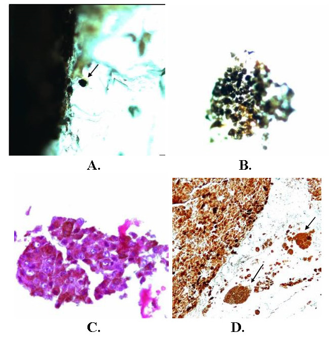
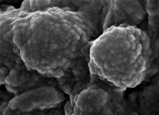
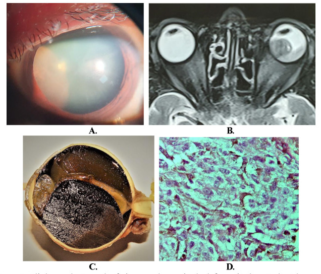
Aim: To report an exceptionally rare case of malignant choroidal melanoma with vitreous seeding, supported by histopathological and field emission scanning electron microscopic (FESEM) studies. Case report: A 58-year-old male with painless diminution of vision in his left eye for past 1 month was found to have a brown retrolental mass lesion on slit lamp examination in the left eye. Detailed fundus examination revealed choroidal melanoma in the left eye with pigmented seeds extending into the vitreous cavity and associated exudative retinal detachment. Ocular imaging was consistent with the diagnosis. Results: The eyeball was enucleated and the tumor was considered as stage IIB (AJCC 8th edition classification). Metastatic workup of the patient was negative. One half of the eyeball was subjected to field emission scanning electron microscopy to further study the nature and appearance of vitreous seeds. Discussion: Vitreous seeding in choroidal melanoma has been reported only in a handful of cases in literature. Histopathological confirmation of vitreous seeds was done in our case and morphological detailing was performed using FESEM study. Conclusions: Treatment naïve choroidal melanoma can very rarely have vitreous seeds. Early enucleation in such cases carries a favorable prognosis.


 分享
分享

 求助内容:
求助内容: 应助结果提醒方式:
 扫码关注我们
