Junming Zhang, Li Liu, Yuexia Li, Jinglei Wu, Xiangxin Lou
{"title":"小鼠胚胎成纤维细胞来源的细胞外基质促进内耳来源细胞的扩增。","authors":"Junming Zhang, Li Liu, Yuexia Li, Jinglei Wu, Xiangxin Lou","doi":"10.22074/cellj.2023.1989426.1228","DOIUrl":null,"url":null,"abstract":"<p><strong>Objective: </strong>Previous reports showed that mouse embryonic fibroblasts (MEFs) could support pluripotent stem cell selfrenewal and maintain their pluripotency. The goal of this study was to reveal whether the decellularized extracellular matrix derived from MEFs (MEF-ECM) is beneficial to promote the proliferation of inner ear-derived cells.</p><p><strong>Materials and methods: </strong>In this experimental study, we prepared a cell-free MEF-ECM through decellularization. Scanning electron microscope (SEM) and immunofluorescent staining were conducted for phenotype characterization. Organs of Corti were dissected from postnatal day 2 and the inner ear-derived cells were obtained. The identification of inner ear-derived cells was conducted by using reverse transcription-polymerase chain reaction (RT-PCR). Cell counting kit-8 (CCK-8) was used to evaluate the proliferation capability of inner ear-derived cells cultured on the MEFECM and tissue culture plate (TCP).</p><p><strong>Results: </strong>The MEF-ECM was clearly observed after decellularization via SEM, and the immunofluorescence staining results revealed that MEF-ECM was composed of three proteins, including collagen I, fibronectin and laminin. Most importantly, the results of CCK-8 showed that compared with TCP, MEF-ECM could effectively facilitate the proliferation of inner ear-derived cells.</p><p><strong>Conclusion: </strong>The discovery of the potential of MEF-ECM in promoting inner ear-derived cell proliferation indicates that the decellularized matrix microenvironment may play a vital role in keeping proliferation ability of these cells. Our findings indicate that the use of MEF-ECM may serve as a novel approach for expanding inner ear-derived cells and potentially facilitating the clinical application of inner ear-derived cells for hearing loss in the future.</p>","PeriodicalId":49224,"journal":{"name":"Cell Journal","volume":"25 7","pages":"447-454"},"PeriodicalIF":1.7000,"publicationDate":"2023-07-25","publicationTypes":"Journal Article","fieldsOfStudy":null,"isOpenAccess":false,"openAccessPdf":"https://ftp.ncbi.nlm.nih.gov/pub/pmc/oa_pdf/b0/e5/Cell-J-25-447.PMC10404357.pdf","citationCount":"0","resultStr":"{\"title\":\"Mouse Embryonic Fibroblasts-Derived Extracellular Matrix Facilitates Expansion of Inner Ear-Derived Cells.\",\"authors\":\"Junming Zhang, Li Liu, Yuexia Li, Jinglei Wu, Xiangxin Lou\",\"doi\":\"10.22074/cellj.2023.1989426.1228\",\"DOIUrl\":null,\"url\":null,\"abstract\":\"<p><strong>Objective: </strong>Previous reports showed that mouse embryonic fibroblasts (MEFs) could support pluripotent stem cell selfrenewal and maintain their pluripotency. The goal of this study was to reveal whether the decellularized extracellular matrix derived from MEFs (MEF-ECM) is beneficial to promote the proliferation of inner ear-derived cells.</p><p><strong>Materials and methods: </strong>In this experimental study, we prepared a cell-free MEF-ECM through decellularization. Scanning electron microscope (SEM) and immunofluorescent staining were conducted for phenotype characterization. Organs of Corti were dissected from postnatal day 2 and the inner ear-derived cells were obtained. The identification of inner ear-derived cells was conducted by using reverse transcription-polymerase chain reaction (RT-PCR). Cell counting kit-8 (CCK-8) was used to evaluate the proliferation capability of inner ear-derived cells cultured on the MEFECM and tissue culture plate (TCP).</p><p><strong>Results: </strong>The MEF-ECM was clearly observed after decellularization via SEM, and the immunofluorescence staining results revealed that MEF-ECM was composed of three proteins, including collagen I, fibronectin and laminin. Most importantly, the results of CCK-8 showed that compared with TCP, MEF-ECM could effectively facilitate the proliferation of inner ear-derived cells.</p><p><strong>Conclusion: </strong>The discovery of the potential of MEF-ECM in promoting inner ear-derived cell proliferation indicates that the decellularized matrix microenvironment may play a vital role in keeping proliferation ability of these cells. Our findings indicate that the use of MEF-ECM may serve as a novel approach for expanding inner ear-derived cells and potentially facilitating the clinical application of inner ear-derived cells for hearing loss in the future.</p>\",\"PeriodicalId\":49224,\"journal\":{\"name\":\"Cell Journal\",\"volume\":\"25 7\",\"pages\":\"447-454\"},\"PeriodicalIF\":1.7000,\"publicationDate\":\"2023-07-25\",\"publicationTypes\":\"Journal Article\",\"fieldsOfStudy\":null,\"isOpenAccess\":false,\"openAccessPdf\":\"https://ftp.ncbi.nlm.nih.gov/pub/pmc/oa_pdf/b0/e5/Cell-J-25-447.PMC10404357.pdf\",\"citationCount\":\"0\",\"resultStr\":null,\"platform\":\"Semanticscholar\",\"paperid\":null,\"PeriodicalName\":\"Cell Journal\",\"FirstCategoryId\":\"99\",\"ListUrlMain\":\"https://doi.org/10.22074/cellj.2023.1989426.1228\",\"RegionNum\":4,\"RegionCategory\":\"生物学\",\"ArticlePicture\":[],\"TitleCN\":null,\"AbstractTextCN\":null,\"PMCID\":null,\"EPubDate\":\"\",\"PubModel\":\"\",\"JCR\":\"Q4\",\"JCRName\":\"CELL BIOLOGY\",\"Score\":null,\"Total\":0}","platform":"Semanticscholar","paperid":null,"PeriodicalName":"Cell Journal","FirstCategoryId":"99","ListUrlMain":"https://doi.org/10.22074/cellj.2023.1989426.1228","RegionNum":4,"RegionCategory":"生物学","ArticlePicture":[],"TitleCN":null,"AbstractTextCN":null,"PMCID":null,"EPubDate":"","PubModel":"","JCR":"Q4","JCRName":"CELL BIOLOGY","Score":null,"Total":0}
Objective: Previous reports showed that mouse embryonic fibroblasts (MEFs) could support pluripotent stem cell selfrenewal and maintain their pluripotency. The goal of this study was to reveal whether the decellularized extracellular matrix derived from MEFs (MEF-ECM) is beneficial to promote the proliferation of inner ear-derived cells.
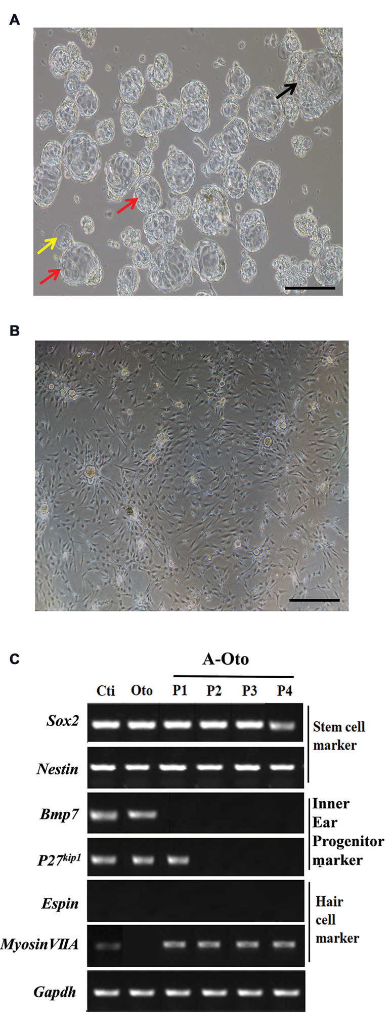
Materials and methods: In this experimental study, we prepared a cell-free MEF-ECM through decellularization. Scanning electron microscope (SEM) and immunofluorescent staining were conducted for phenotype characterization. Organs of Corti were dissected from postnatal day 2 and the inner ear-derived cells were obtained. The identification of inner ear-derived cells was conducted by using reverse transcription-polymerase chain reaction (RT-PCR). Cell counting kit-8 (CCK-8) was used to evaluate the proliferation capability of inner ear-derived cells cultured on the MEFECM and tissue culture plate (TCP).
Results: The MEF-ECM was clearly observed after decellularization via SEM, and the immunofluorescence staining results revealed that MEF-ECM was composed of three proteins, including collagen I, fibronectin and laminin. Most importantly, the results of CCK-8 showed that compared with TCP, MEF-ECM could effectively facilitate the proliferation of inner ear-derived cells.
Conclusion: The discovery of the potential of MEF-ECM in promoting inner ear-derived cell proliferation indicates that the decellularized matrix microenvironment may play a vital role in keeping proliferation ability of these cells. Our findings indicate that the use of MEF-ECM may serve as a novel approach for expanding inner ear-derived cells and potentially facilitating the clinical application of inner ear-derived cells for hearing loss in the future.
期刊介绍:
The “Cell Journal (Yakhteh)“, formerly published as “Yakhteh Medical Journal”, is a quarterly English publication of Royan Institute. This journal focuses on topics relevant to cellular and molecular scientific areas, besides other related fields. The Cell J has been certified by Ministry of Culture and Islamic Guidance in 1999 and was accredited as a scientific and research journal by HBI (Health and Biomedical Information) Journal Accreditation Commission in 2000 which is an open access journal.


 分享
分享
 求助内容:
求助内容: 应助结果提醒方式:
 扫码关注我们
