Yi Dong, Wen-Ping Wang, Andre Ignee, Dan Zuo, Yi-Jie Qiu, Qi Zhang, Xiu-Yun Lu, Sheng Chen, Christoph Frank Dietrich
{"title":"多普勒电阻指数在局灶性肝病变鉴别诊断中的价值。","authors":"Yi Dong, Wen-Ping Wang, Andre Ignee, Dan Zuo, Yi-Jie Qiu, Qi Zhang, Xiu-Yun Lu, Sheng Chen, Christoph Frank Dietrich","doi":"10.15557/jou.2023.0010","DOIUrl":null,"url":null,"abstract":"<p><strong>Aim: </strong>To investigate the diagnostic value of resistance index (RI) in differentiating focal liver lesions.</p><p><strong>Patients and methods: </strong>In this retrospective study, a total of 576 patients with histologically confirmed focal liver lesions were included. Each patient underwent B-mode ultrasound examination and color Doppler ultrasound examination. The RI values of different focal liver lesions were recorded and compared.</p><p><strong>Results: </strong>The mean RI value of benign lesions was significantly lower than that of malignant lesions (0.54 ± 0.10 <i>vs</i>. 0.71 ± 0.12) (<i>p</i> <0.05). In malignant lesions, the RI value of intrahepatic cholangiocarcinoma was significantly lower than that of hepatocellular carcinoma lesions. Furthermore, in hepatocellular carcinoma lesions, the RI of large lesions (group 4: >10 cm) was significantly lower than that of small lesions (group 1: ≤2 cm, group 2: 2-5 cm) (<i>p</i> <0.05). Taken RI of 0.615 as a cutoff value to differentiate malignant and benign lesions, the sensitivity, specificity, positive predictive value and negative predictive value were 82.80%, 81.00%, 81.34% and 82.48%, respectively.</p><p><strong>Conclusion: </strong>Color Doppler ultrasound examination is a valuable imaging method in detecting blood flow signal within liver lesions. The RI parameter should be helpful in differentiating malignant and benign liver tumors.</p>","PeriodicalId":45612,"journal":{"name":"Journal of Ultrasonography","volume":"23 93","pages":"e45-e52"},"PeriodicalIF":1.5000,"publicationDate":"2023-06-01","publicationTypes":"Journal Article","fieldsOfStudy":null,"isOpenAccess":false,"openAccessPdf":"https://ftp.ncbi.nlm.nih.gov/pub/pmc/oa_pdf/11/a6/jou-23-93-jou.2023.0010.PMC10379844.pdf","citationCount":"2","resultStr":"{\"title\":\"The diagnostic value of Doppler Resistive Index in the differential diagnosis of focal liver lesions.\",\"authors\":\"Yi Dong, Wen-Ping Wang, Andre Ignee, Dan Zuo, Yi-Jie Qiu, Qi Zhang, Xiu-Yun Lu, Sheng Chen, Christoph Frank Dietrich\",\"doi\":\"10.15557/jou.2023.0010\",\"DOIUrl\":null,\"url\":null,\"abstract\":\"<p><strong>Aim: </strong>To investigate the diagnostic value of resistance index (RI) in differentiating focal liver lesions.</p><p><strong>Patients and methods: </strong>In this retrospective study, a total of 576 patients with histologically confirmed focal liver lesions were included. Each patient underwent B-mode ultrasound examination and color Doppler ultrasound examination. The RI values of different focal liver lesions were recorded and compared.</p><p><strong>Results: </strong>The mean RI value of benign lesions was significantly lower than that of malignant lesions (0.54 ± 0.10 <i>vs</i>. 0.71 ± 0.12) (<i>p</i> <0.05). In malignant lesions, the RI value of intrahepatic cholangiocarcinoma was significantly lower than that of hepatocellular carcinoma lesions. Furthermore, in hepatocellular carcinoma lesions, the RI of large lesions (group 4: >10 cm) was significantly lower than that of small lesions (group 1: ≤2 cm, group 2: 2-5 cm) (<i>p</i> <0.05). Taken RI of 0.615 as a cutoff value to differentiate malignant and benign lesions, the sensitivity, specificity, positive predictive value and negative predictive value were 82.80%, 81.00%, 81.34% and 82.48%, respectively.</p><p><strong>Conclusion: </strong>Color Doppler ultrasound examination is a valuable imaging method in detecting blood flow signal within liver lesions. The RI parameter should be helpful in differentiating malignant and benign liver tumors.</p>\",\"PeriodicalId\":45612,\"journal\":{\"name\":\"Journal of Ultrasonography\",\"volume\":\"23 93\",\"pages\":\"e45-e52\"},\"PeriodicalIF\":1.5000,\"publicationDate\":\"2023-06-01\",\"publicationTypes\":\"Journal Article\",\"fieldsOfStudy\":null,\"isOpenAccess\":false,\"openAccessPdf\":\"https://ftp.ncbi.nlm.nih.gov/pub/pmc/oa_pdf/11/a6/jou-23-93-jou.2023.0010.PMC10379844.pdf\",\"citationCount\":\"2\",\"resultStr\":null,\"platform\":\"Semanticscholar\",\"paperid\":null,\"PeriodicalName\":\"Journal of Ultrasonography\",\"FirstCategoryId\":\"1085\",\"ListUrlMain\":\"https://doi.org/10.15557/jou.2023.0010\",\"RegionNum\":0,\"RegionCategory\":null,\"ArticlePicture\":[],\"TitleCN\":null,\"AbstractTextCN\":null,\"PMCID\":null,\"EPubDate\":\"\",\"PubModel\":\"\",\"JCR\":\"Q3\",\"JCRName\":\"RADIOLOGY, NUCLEAR MEDICINE & MEDICAL IMAGING\",\"Score\":null,\"Total\":0}","platform":"Semanticscholar","paperid":null,"PeriodicalName":"Journal of Ultrasonography","FirstCategoryId":"1085","ListUrlMain":"https://doi.org/10.15557/jou.2023.0010","RegionNum":0,"RegionCategory":null,"ArticlePicture":[],"TitleCN":null,"AbstractTextCN":null,"PMCID":null,"EPubDate":"","PubModel":"","JCR":"Q3","JCRName":"RADIOLOGY, NUCLEAR MEDICINE & MEDICAL IMAGING","Score":null,"Total":0}
引用次数: 2
摘要
目的:探讨耐药指数(RI)对肝局灶性病变的诊断价值。患者和方法:本回顾性研究共纳入576例组织学证实的局灶性肝脏病变患者。分别行b超和彩色多普勒超声检查。记录不同局灶性肝病变的RI值并进行比较。结果:良性病变的平均RI值显著低于恶性病变(0.54±0.10 vs 0.71±0.12)(p 10 cm),显著低于小病变(1组≤2 cm, 2组2 ~ 5 cm) (p结论:彩色多普勒超声检查是检测肝脏病变内血流信号的一种有价值的影像学方法。RI参数应有助于鉴别肝肿瘤的良恶性。
The diagnostic value of Doppler Resistive Index in the differential diagnosis of focal liver lesions.
Aim: To investigate the diagnostic value of resistance index (RI) in differentiating focal liver lesions.
Patients and methods: In this retrospective study, a total of 576 patients with histologically confirmed focal liver lesions were included. Each patient underwent B-mode ultrasound examination and color Doppler ultrasound examination. The RI values of different focal liver lesions were recorded and compared.
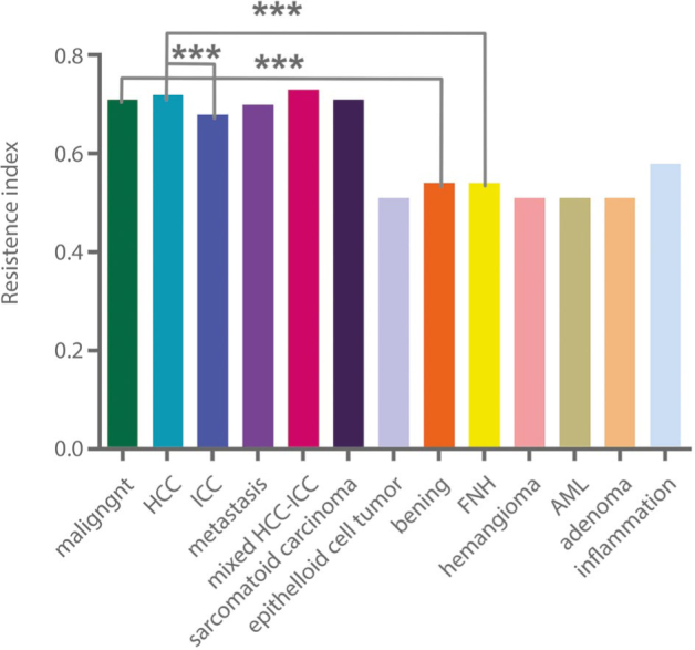
Results: The mean RI value of benign lesions was significantly lower than that of malignant lesions (0.54 ± 0.10 vs. 0.71 ± 0.12) (p <0.05). In malignant lesions, the RI value of intrahepatic cholangiocarcinoma was significantly lower than that of hepatocellular carcinoma lesions. Furthermore, in hepatocellular carcinoma lesions, the RI of large lesions (group 4: >10 cm) was significantly lower than that of small lesions (group 1: ≤2 cm, group 2: 2-5 cm) (p <0.05). Taken RI of 0.615 as a cutoff value to differentiate malignant and benign lesions, the sensitivity, specificity, positive predictive value and negative predictive value were 82.80%, 81.00%, 81.34% and 82.48%, respectively.
Conclusion: Color Doppler ultrasound examination is a valuable imaging method in detecting blood flow signal within liver lesions. The RI parameter should be helpful in differentiating malignant and benign liver tumors.


 分享
分享
 求助内容:
求助内容: 应助结果提醒方式:
 扫码关注我们
