Seth B Boren, Sean I Savitz, Nicole Gonzales, Khader Hasan, Andrea Becerril-Gaitan, Vahed Maroufy, Yuan Li, James Grotta, Emily A Steven, Ching-Jen Chen, Clark W Sitton, Jaroslaw Aronowski, Muhammad E Haque
{"title":"出血性脑卒中后皮质脊髓束形状的纵向形态变化","authors":"Seth B Boren, Sean I Savitz, Nicole Gonzales, Khader Hasan, Andrea Becerril-Gaitan, Vahed Maroufy, Yuan Li, James Grotta, Emily A Steven, Ching-Jen Chen, Clark W Sitton, Jaroslaw Aronowski, Muhammad E Haque","doi":"10.1007/s12975-023-01168-y","DOIUrl":null,"url":null,"abstract":"<p><p>Deep intracerebral hemorrhage (ICH) exerts a direct force on corticospinal tracts (CST) causing shape deformation. Using serial MRI, Generalized Procrustes Analysis (GPA), and Principal Components Analysis (PCA), we temporally evaluated the change in CST shape. Thirty-five deep ICH patients with ipsilesional-CST deformation were serially imaged on a 3T-MRI with a median imaging time of day-2 and 84 of onset. Anatomical and diffusion tensor images (DTI) were acquired. Using DTI color-coded maps, 15 landmarks were drawn on each CST and the centroids were computed in 3 dimensions. The contralesional-CST landmarks were used as a reference. The GPA outlined the shape coordinates and we superimposed the ipsilesional-CST shape at the two-time points. A multivariate PCA was applied to identify eigenvectors associated with the highest percentile of change. The first three principal components representing CST deformation along the left-right (PC1), anterior-posterior (PC2), and superior-inferior (PC3) respectively were responsible for 57.9% of shape variance. The PC1 (36.1%, p < 0.0001) and PC3 (9.58%, p < 0.01) showed a significant deformation between the two-time points. Compared to the contralesional-CST, the ipsilesional PC scores were significantly (p < 0.0001) different only at the first-timepoint. A significant positive association between the ipsilesional-CST deformation and hematoma volume was observed. We present a novel method to quantify CST deformation caused by ICH. Deformation most often occurs in left-right axis (PC1) and superior-inferior (PC3) directions. As compared to the reference, the significant temporal difference at the first time point suggests CST restoration over time.</p>","PeriodicalId":23237,"journal":{"name":"Translational Stroke Research","volume":" ","pages":"893-901"},"PeriodicalIF":3.8000,"publicationDate":"2024-10-01","publicationTypes":"Journal Article","fieldsOfStudy":null,"isOpenAccess":false,"openAccessPdf":"","citationCount":"0","resultStr":"{\"title\":\"Longitudinal Morphometric Changes in the Corticospinal Tract Shape After Hemorrhagic Stroke.\",\"authors\":\"Seth B Boren, Sean I Savitz, Nicole Gonzales, Khader Hasan, Andrea Becerril-Gaitan, Vahed Maroufy, Yuan Li, James Grotta, Emily A Steven, Ching-Jen Chen, Clark W Sitton, Jaroslaw Aronowski, Muhammad E Haque\",\"doi\":\"10.1007/s12975-023-01168-y\",\"DOIUrl\":null,\"url\":null,\"abstract\":\"<p><p>Deep intracerebral hemorrhage (ICH) exerts a direct force on corticospinal tracts (CST) causing shape deformation. Using serial MRI, Generalized Procrustes Analysis (GPA), and Principal Components Analysis (PCA), we temporally evaluated the change in CST shape. Thirty-five deep ICH patients with ipsilesional-CST deformation were serially imaged on a 3T-MRI with a median imaging time of day-2 and 84 of onset. Anatomical and diffusion tensor images (DTI) were acquired. Using DTI color-coded maps, 15 landmarks were drawn on each CST and the centroids were computed in 3 dimensions. The contralesional-CST landmarks were used as a reference. The GPA outlined the shape coordinates and we superimposed the ipsilesional-CST shape at the two-time points. A multivariate PCA was applied to identify eigenvectors associated with the highest percentile of change. The first three principal components representing CST deformation along the left-right (PC1), anterior-posterior (PC2), and superior-inferior (PC3) respectively were responsible for 57.9% of shape variance. The PC1 (36.1%, p < 0.0001) and PC3 (9.58%, p < 0.01) showed a significant deformation between the two-time points. Compared to the contralesional-CST, the ipsilesional PC scores were significantly (p < 0.0001) different only at the first-timepoint. A significant positive association between the ipsilesional-CST deformation and hematoma volume was observed. We present a novel method to quantify CST deformation caused by ICH. Deformation most often occurs in left-right axis (PC1) and superior-inferior (PC3) directions. As compared to the reference, the significant temporal difference at the first time point suggests CST restoration over time.</p>\",\"PeriodicalId\":23237,\"journal\":{\"name\":\"Translational Stroke Research\",\"volume\":\" \",\"pages\":\"893-901\"},\"PeriodicalIF\":3.8000,\"publicationDate\":\"2024-10-01\",\"publicationTypes\":\"Journal Article\",\"fieldsOfStudy\":null,\"isOpenAccess\":false,\"openAccessPdf\":\"\",\"citationCount\":\"0\",\"resultStr\":null,\"platform\":\"Semanticscholar\",\"paperid\":null,\"PeriodicalName\":\"Translational Stroke Research\",\"FirstCategoryId\":\"3\",\"ListUrlMain\":\"https://doi.org/10.1007/s12975-023-01168-y\",\"RegionNum\":2,\"RegionCategory\":\"医学\",\"ArticlePicture\":[],\"TitleCN\":null,\"AbstractTextCN\":null,\"PMCID\":null,\"EPubDate\":\"2023/6/13 0:00:00\",\"PubModel\":\"Epub\",\"JCR\":\"Q1\",\"JCRName\":\"CLINICAL NEUROLOGY\",\"Score\":null,\"Total\":0}","platform":"Semanticscholar","paperid":null,"PeriodicalName":"Translational Stroke Research","FirstCategoryId":"3","ListUrlMain":"https://doi.org/10.1007/s12975-023-01168-y","RegionNum":2,"RegionCategory":"医学","ArticlePicture":[],"TitleCN":null,"AbstractTextCN":null,"PMCID":null,"EPubDate":"2023/6/13 0:00:00","PubModel":"Epub","JCR":"Q1","JCRName":"CLINICAL NEUROLOGY","Score":null,"Total":0}
Longitudinal Morphometric Changes in the Corticospinal Tract Shape After Hemorrhagic Stroke.
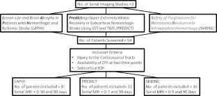
Deep intracerebral hemorrhage (ICH) exerts a direct force on corticospinal tracts (CST) causing shape deformation. Using serial MRI, Generalized Procrustes Analysis (GPA), and Principal Components Analysis (PCA), we temporally evaluated the change in CST shape. Thirty-five deep ICH patients with ipsilesional-CST deformation were serially imaged on a 3T-MRI with a median imaging time of day-2 and 84 of onset. Anatomical and diffusion tensor images (DTI) were acquired. Using DTI color-coded maps, 15 landmarks were drawn on each CST and the centroids were computed in 3 dimensions. The contralesional-CST landmarks were used as a reference. The GPA outlined the shape coordinates and we superimposed the ipsilesional-CST shape at the two-time points. A multivariate PCA was applied to identify eigenvectors associated with the highest percentile of change. The first three principal components representing CST deformation along the left-right (PC1), anterior-posterior (PC2), and superior-inferior (PC3) respectively were responsible for 57.9% of shape variance. The PC1 (36.1%, p < 0.0001) and PC3 (9.58%, p < 0.01) showed a significant deformation between the two-time points. Compared to the contralesional-CST, the ipsilesional PC scores were significantly (p < 0.0001) different only at the first-timepoint. A significant positive association between the ipsilesional-CST deformation and hematoma volume was observed. We present a novel method to quantify CST deformation caused by ICH. Deformation most often occurs in left-right axis (PC1) and superior-inferior (PC3) directions. As compared to the reference, the significant temporal difference at the first time point suggests CST restoration over time.
期刊介绍:
Translational Stroke Research covers basic, translational, and clinical studies. The Journal emphasizes novel approaches to help both to understand clinical phenomenon through basic science tools, and to translate basic science discoveries into the development of new strategies for the prevention, assessment, treatment, and enhancement of central nervous system repair after stroke and other forms of neurotrauma.
Translational Stroke Research focuses on translational research and is relevant to both basic scientists and physicians, including but not restricted to neuroscientists, vascular biologists, neurologists, neuroimagers, and neurosurgeons.


 分享
分享
 求助内容:
求助内容: 应助结果提醒方式:
 扫码关注我们
