Vendhan Ramanujam, Lee Tian, Clarence Chow, Mark C Kendall
{"title":"常用外围块的三维成像:使用手持式护理点超声系统。","authors":"Vendhan Ramanujam, Lee Tian, Clarence Chow, Mark C Kendall","doi":"10.5812/aapm-134797","DOIUrl":null,"url":null,"abstract":"<p><strong>Background: </strong>Handheld ultrasound devices have become popular among clinicians due to their affordability and compatibility with tablets and smartphones. Several handheld ultrasound devices have the capability to construct three-dimensional (3D) images using a traditional two-dimensional (2D) ultrasound transducer.</p><p><strong>Objectives: </strong>The current study aimed to construct 3D images of common peripheral nerve and fascial plane blocks using a handheld ultrasound device with a 2D ultrasound probe.</p><p><strong>Methods: </strong>A total of 10 patients who were scheduled to receive ultrasound-guided peripheral nerve blocks for outpatient surgery and classified as the American Society of Anesthesiologists physical status I or II with a body mass index of ≤ 30 kg/m<sup>2</sup> were included in the study. Patients who presented with anatomical variations during the initial ultrasound scanning were excluded.</p><p><strong>Results: </strong>This study successfully constructed 3D images of 10 peripheral nerve blocks. The average time to complete each 3D scan was less than 5 seconds per attempt, with fascial plane blocks requiring twice the amount of time to complete. All the nerve blocks provided effective postoperative analgesia without complications. The 3D images were successfully captured in all patients.</p><p><strong>Conclusions: </strong>The 3D images provide clinicians with valuable information on the anatomical boundaries of the injectate that can further direct needle direction and placement of local anesthetic to achieve visual confidence of anesthetic spread.</p>","PeriodicalId":7841,"journal":{"name":"Anesthesiology and Pain Medicine","volume":"13 2","pages":"e134797"},"PeriodicalIF":0.0000,"publicationDate":"2023-04-01","publicationTypes":"Journal Article","fieldsOfStudy":null,"isOpenAccess":false,"openAccessPdf":"https://ftp.ncbi.nlm.nih.gov/pub/pmc/oa_pdf/9f/24/aapm-13-2-134797.PMC10389033.pdf","citationCount":"0","resultStr":"{\"title\":\"Three-Dimensional Imaging of Commonly Performed Peripheral Blocks: Using a Handheld Point-of-Care Ultrasound System.\",\"authors\":\"Vendhan Ramanujam, Lee Tian, Clarence Chow, Mark C Kendall\",\"doi\":\"10.5812/aapm-134797\",\"DOIUrl\":null,\"url\":null,\"abstract\":\"<p><strong>Background: </strong>Handheld ultrasound devices have become popular among clinicians due to their affordability and compatibility with tablets and smartphones. Several handheld ultrasound devices have the capability to construct three-dimensional (3D) images using a traditional two-dimensional (2D) ultrasound transducer.</p><p><strong>Objectives: </strong>The current study aimed to construct 3D images of common peripheral nerve and fascial plane blocks using a handheld ultrasound device with a 2D ultrasound probe.</p><p><strong>Methods: </strong>A total of 10 patients who were scheduled to receive ultrasound-guided peripheral nerve blocks for outpatient surgery and classified as the American Society of Anesthesiologists physical status I or II with a body mass index of ≤ 30 kg/m<sup>2</sup> were included in the study. Patients who presented with anatomical variations during the initial ultrasound scanning were excluded.</p><p><strong>Results: </strong>This study successfully constructed 3D images of 10 peripheral nerve blocks. The average time to complete each 3D scan was less than 5 seconds per attempt, with fascial plane blocks requiring twice the amount of time to complete. All the nerve blocks provided effective postoperative analgesia without complications. The 3D images were successfully captured in all patients.</p><p><strong>Conclusions: </strong>The 3D images provide clinicians with valuable information on the anatomical boundaries of the injectate that can further direct needle direction and placement of local anesthetic to achieve visual confidence of anesthetic spread.</p>\",\"PeriodicalId\":7841,\"journal\":{\"name\":\"Anesthesiology and Pain Medicine\",\"volume\":\"13 2\",\"pages\":\"e134797\"},\"PeriodicalIF\":0.0000,\"publicationDate\":\"2023-04-01\",\"publicationTypes\":\"Journal Article\",\"fieldsOfStudy\":null,\"isOpenAccess\":false,\"openAccessPdf\":\"https://ftp.ncbi.nlm.nih.gov/pub/pmc/oa_pdf/9f/24/aapm-13-2-134797.PMC10389033.pdf\",\"citationCount\":\"0\",\"resultStr\":null,\"platform\":\"Semanticscholar\",\"paperid\":null,\"PeriodicalName\":\"Anesthesiology and Pain Medicine\",\"FirstCategoryId\":\"1085\",\"ListUrlMain\":\"https://doi.org/10.5812/aapm-134797\",\"RegionNum\":0,\"RegionCategory\":null,\"ArticlePicture\":[],\"TitleCN\":null,\"AbstractTextCN\":null,\"PMCID\":null,\"EPubDate\":\"\",\"PubModel\":\"\",\"JCR\":\"Q2\",\"JCRName\":\"Medicine\",\"Score\":null,\"Total\":0}","platform":"Semanticscholar","paperid":null,"PeriodicalName":"Anesthesiology and Pain Medicine","FirstCategoryId":"1085","ListUrlMain":"https://doi.org/10.5812/aapm-134797","RegionNum":0,"RegionCategory":null,"ArticlePicture":[],"TitleCN":null,"AbstractTextCN":null,"PMCID":null,"EPubDate":"","PubModel":"","JCR":"Q2","JCRName":"Medicine","Score":null,"Total":0}
Three-Dimensional Imaging of Commonly Performed Peripheral Blocks: Using a Handheld Point-of-Care Ultrasound System.
Background: Handheld ultrasound devices have become popular among clinicians due to their affordability and compatibility with tablets and smartphones. Several handheld ultrasound devices have the capability to construct three-dimensional (3D) images using a traditional two-dimensional (2D) ultrasound transducer.
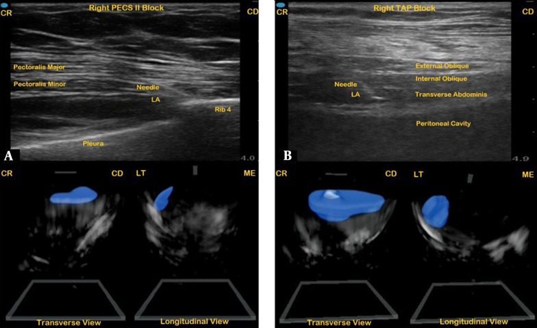
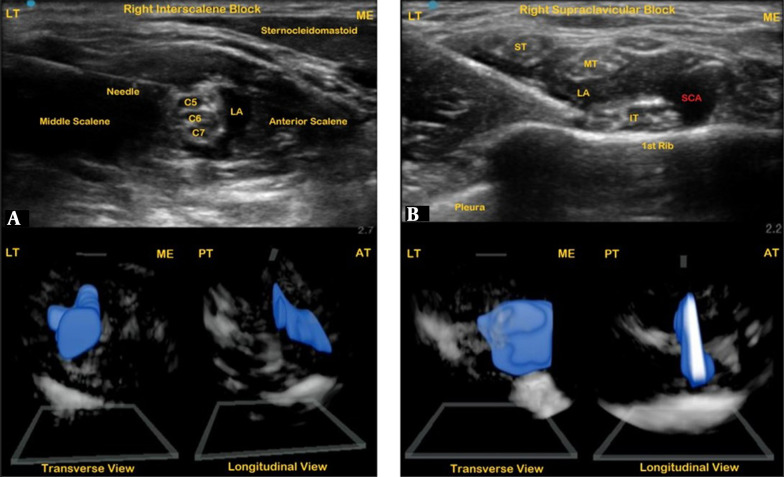
Objectives: The current study aimed to construct 3D images of common peripheral nerve and fascial plane blocks using a handheld ultrasound device with a 2D ultrasound probe.
Methods: A total of 10 patients who were scheduled to receive ultrasound-guided peripheral nerve blocks for outpatient surgery and classified as the American Society of Anesthesiologists physical status I or II with a body mass index of ≤ 30 kg/m2 were included in the study. Patients who presented with anatomical variations during the initial ultrasound scanning were excluded.
Results: This study successfully constructed 3D images of 10 peripheral nerve blocks. The average time to complete each 3D scan was less than 5 seconds per attempt, with fascial plane blocks requiring twice the amount of time to complete. All the nerve blocks provided effective postoperative analgesia without complications. The 3D images were successfully captured in all patients.
Conclusions: The 3D images provide clinicians with valuable information on the anatomical boundaries of the injectate that can further direct needle direction and placement of local anesthetic to achieve visual confidence of anesthetic spread.


 分享
分享
 求助内容:
求助内容: 应助结果提醒方式:
 扫码关注我们
