Molly Tzu-Yu Lin, Isabelle Xin Yu Lee, Wei-Li Chen, Mei-Yun Chen, Jodhbir S Mehta, Gary H F Yam, Gary S L Peh, Yu-Chi Liu
{"title":"Culture of Primary Neurons from Dissociated and Cryopreserved Mouse Trigeminal Ganglion.","authors":"Molly Tzu-Yu Lin, Isabelle Xin Yu Lee, Wei-Li Chen, Mei-Yun Chen, Jodhbir S Mehta, Gary H F Yam, Gary S L Peh, Yu-Chi Liu","doi":"10.1089/ten.TEC.2023.0054","DOIUrl":null,"url":null,"abstract":"<p><p>Corneal nerves originate from the ophthalmic branch of the trigeminal nerve, which enters the cornea at the limbus radially from all directions toward the central cornea. The cell bodies of the sensory neurons of trigeminal nerve are located in the trigeminal ganglion (TG), while the axons are extended into the three divisions, including ophthalmic branch that supplies corneal nerves. Study of primary neuronal cultures established from the TG fibers can therefore provide a knowledge basis for corneal nerve biology and potentially be developed as an <i>in vitro</i> platform for drug testing. However, setting up primary neuron cultures from animal TG has been dubious with inconsistency among laboratories due to a lack of efficient isolation protocol, resulting in low yield and heterogenous cultures. In this study, we used a combined enzymatic digestion with collagenase and TrypLE to dissociate mouse TG while preserving nerve cell viability. A subsequent discontinuous Percoll density gradient followed by mitotic inhibitor treatment effectively diminished the contamination of non-neuronal cells. Using this method, we reproducibly generated high yield and homogenous primary TG neuron cultures. Similar efficiency of nerve cell isolation and culture was further obtained for TG tissue cryopreserved for short (1 week) and long duration (3 months), compared to freshly isolated tissues. In conclusion, this optimized protocol shows a promising potential to standardize TG nerve culture and generate a high-quality corneal nerve model for drug testing and neurotoxicity studies.</p>","PeriodicalId":23154,"journal":{"name":"Tissue engineering. Part C, Methods","volume":"29 8","pages":"381-393"},"PeriodicalIF":2.6000,"publicationDate":"2023-08-01","publicationTypes":"Journal Article","fieldsOfStudy":null,"isOpenAccess":false,"openAccessPdf":"https://www.ncbi.nlm.nih.gov/pmc/articles/PMC10442681/pdf/","citationCount":"0","resultStr":null,"platform":"Semanticscholar","paperid":null,"PeriodicalName":"Tissue engineering. Part C, Methods","FirstCategoryId":"3","ListUrlMain":"https://doi.org/10.1089/ten.TEC.2023.0054","RegionNum":4,"RegionCategory":"医学","ArticlePicture":[],"TitleCN":null,"AbstractTextCN":null,"PMCID":null,"EPubDate":"","PubModel":"","JCR":"Q3","JCRName":"CELL & TISSUE ENGINEERING","Score":null,"Total":0}
引用次数: 0
Abstract
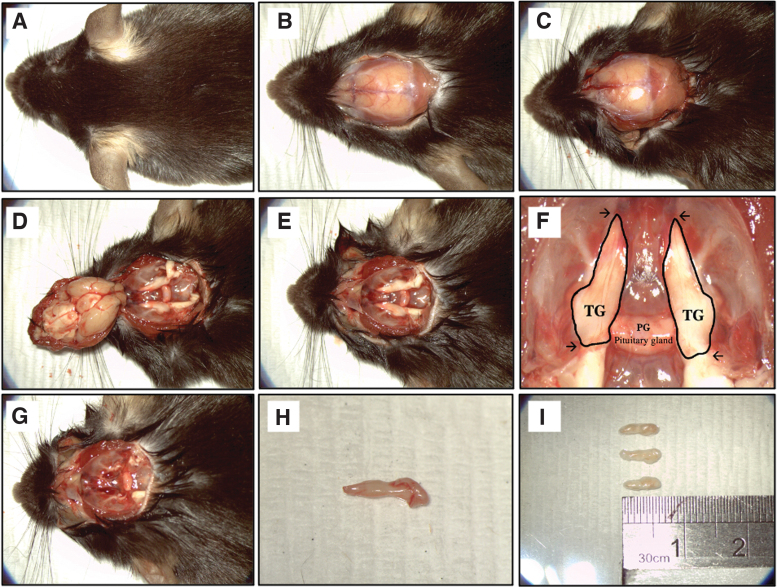
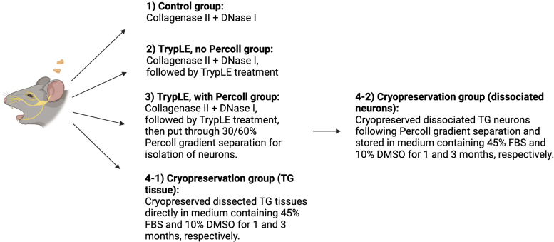
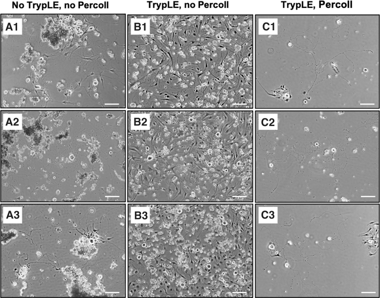
Corneal nerves originate from the ophthalmic branch of the trigeminal nerve, which enters the cornea at the limbus radially from all directions toward the central cornea. The cell bodies of the sensory neurons of trigeminal nerve are located in the trigeminal ganglion (TG), while the axons are extended into the three divisions, including ophthalmic branch that supplies corneal nerves. Study of primary neuronal cultures established from the TG fibers can therefore provide a knowledge basis for corneal nerve biology and potentially be developed as an in vitro platform for drug testing. However, setting up primary neuron cultures from animal TG has been dubious with inconsistency among laboratories due to a lack of efficient isolation protocol, resulting in low yield and heterogenous cultures. In this study, we used a combined enzymatic digestion with collagenase and TrypLE to dissociate mouse TG while preserving nerve cell viability. A subsequent discontinuous Percoll density gradient followed by mitotic inhibitor treatment effectively diminished the contamination of non-neuronal cells. Using this method, we reproducibly generated high yield and homogenous primary TG neuron cultures. Similar efficiency of nerve cell isolation and culture was further obtained for TG tissue cryopreserved for short (1 week) and long duration (3 months), compared to freshly isolated tissues. In conclusion, this optimized protocol shows a promising potential to standardize TG nerve culture and generate a high-quality corneal nerve model for drug testing and neurotoxicity studies.
期刊介绍:
Tissue Engineering is the preeminent, biomedical journal advancing the field with cutting-edge research and applications that repair or regenerate portions or whole tissues. This multidisciplinary journal brings together the principles of engineering and life sciences in the creation of artificial tissues and regenerative medicine. Tissue Engineering is divided into three parts, providing a central forum for groundbreaking scientific research and developments of clinical applications from leading experts in the field that will enable the functional replacement of tissues.
Tissue Engineering Methods (Part C) presents innovative tools and assays in scaffold development, stem cells and biologically active molecules to advance the field and to support clinical translation. Part C publishes monthly.


 分享
分享
 求助内容:
求助内容: 应助结果提醒方式:
 扫码关注我们
