José S Ponte, Jesús A Pérez-Guerrero, Francisco Aa Aragão, Yasmin At Menezes, Marcelo M Melo, Igor I Castro-Silva
{"title":"Histomorphometric evaluation of human extraction sockets treated with autologous fibrin, sticky bone or biphasic calcium phosphate.","authors":"José S Ponte, Jesús A Pérez-Guerrero, Francisco Aa Aragão, Yasmin At Menezes, Marcelo M Melo, Igor I Castro-Silva","doi":"10.54589/aol.34/3/271","DOIUrl":null,"url":null,"abstract":"<p><p>It is essential to maintain the alveolar bone ridge to ensure the success of implant therapy. Platelet-rich fibrin (PRF) may benefit bone repair, but its quantitative microscopic results are still inconclusive. The aim of this study was to histomorphometrically analyze human dental alveoli after extraction treated with autologous fibrin, biphasic calcium phosphate or sticky bone. The sample consisted of healthy adult volunteer patients, with clinical and tomographic indication for single post-extraction graft of upper premolars for maintenance of the alveolar ridge and subsequent implantation. The 10 remaining patients in the study were divided into three groups according to the type of filling used in the dental socket: autologous PRF plug covered by a PRF membrane (G1), PRF associated with an alloplastic graft based on hydroxyapatite with beta tricalcium phosphate covered by a collagen membrane (G2) or alloplastic graft based on beta tricalcium phosphate covered by collagen membrane (control). After 8 months, bone biopsies were performed at the grafted sites and the patients underwent implant-prosthetic rehabilitation. Paraffin-embedded tissue blocks were routinely processed and sectionsfrom different depths were mounted in 3 slides and stained with HE. The histomorphometric evaluation analyzed 30 photomicrographs per block, quantifying the percentage presence of newly formed bone, connective tissue and remaining biomaterial using the ImageJ software. Parametric data enabled intergroup comparisons using ANOVA and Tukey's post-hoc test for multiple comparison with statistical significance of 5% (p<0.05), with normality of the 3 groups by the Jarque-Bera test (p>0.05). There was a higher mean of newly formed bone in G1 (68.83%) compared to G2 (35.69%) and control (16.28%). There was greater presence of connective tissue in the control (61.56%). Remaining biomaterial was higher in G2 (15.75%), but did not differ statistically from the control. Bone regeneration obtained with PRF alone or sticky bone suggests the efficacy of these therapies, encouraging the clinical use of this blood concentrate in dental procedures.</p>","PeriodicalId":7033,"journal":{"name":"Acta odontologica latinoamericana : AOL","volume":"34 3","pages":"271-281"},"PeriodicalIF":0.0000,"publicationDate":"2021-12-31","publicationTypes":"Journal Article","fieldsOfStudy":null,"isOpenAccess":false,"openAccessPdf":"https://ftp.ncbi.nlm.nih.gov/pub/pmc/oa_pdf/96/b9/1852-4834-34-3-271.PMC10315085.pdf","citationCount":"0","resultStr":null,"platform":"Semanticscholar","paperid":null,"PeriodicalName":"Acta odontologica latinoamericana : AOL","FirstCategoryId":"1085","ListUrlMain":"https://doi.org/10.54589/aol.34/3/271","RegionNum":0,"RegionCategory":null,"ArticlePicture":[],"TitleCN":null,"AbstractTextCN":null,"PMCID":null,"EPubDate":"","PubModel":"","JCR":"","JCRName":"","Score":null,"Total":0}
引用次数: 0
Abstract
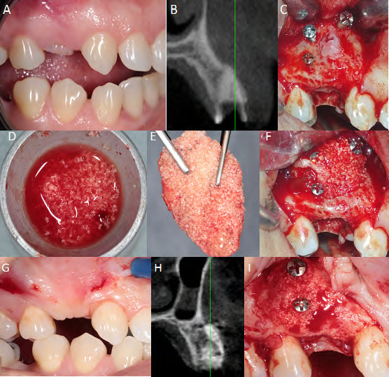
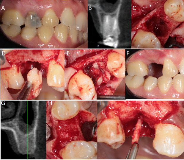
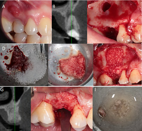
It is essential to maintain the alveolar bone ridge to ensure the success of implant therapy. Platelet-rich fibrin (PRF) may benefit bone repair, but its quantitative microscopic results are still inconclusive. The aim of this study was to histomorphometrically analyze human dental alveoli after extraction treated with autologous fibrin, biphasic calcium phosphate or sticky bone. The sample consisted of healthy adult volunteer patients, with clinical and tomographic indication for single post-extraction graft of upper premolars for maintenance of the alveolar ridge and subsequent implantation. The 10 remaining patients in the study were divided into three groups according to the type of filling used in the dental socket: autologous PRF plug covered by a PRF membrane (G1), PRF associated with an alloplastic graft based on hydroxyapatite with beta tricalcium phosphate covered by a collagen membrane (G2) or alloplastic graft based on beta tricalcium phosphate covered by collagen membrane (control). After 8 months, bone biopsies were performed at the grafted sites and the patients underwent implant-prosthetic rehabilitation. Paraffin-embedded tissue blocks were routinely processed and sectionsfrom different depths were mounted in 3 slides and stained with HE. The histomorphometric evaluation analyzed 30 photomicrographs per block, quantifying the percentage presence of newly formed bone, connective tissue and remaining biomaterial using the ImageJ software. Parametric data enabled intergroup comparisons using ANOVA and Tukey's post-hoc test for multiple comparison with statistical significance of 5% (p<0.05), with normality of the 3 groups by the Jarque-Bera test (p>0.05). There was a higher mean of newly formed bone in G1 (68.83%) compared to G2 (35.69%) and control (16.28%). There was greater presence of connective tissue in the control (61.56%). Remaining biomaterial was higher in G2 (15.75%), but did not differ statistically from the control. Bone regeneration obtained with PRF alone or sticky bone suggests the efficacy of these therapies, encouraging the clinical use of this blood concentrate in dental procedures.


 分享
分享

 求助内容:
求助内容: 应助结果提醒方式:
 扫码关注我们
