Ali Fahd, Ahmed Talaat Temerek, Mohamed T Ellabban, Samar Ahmed Nouby Adam, Sarah Diaa Abd El-Wahab Shaheen, Mervat S Refai, Zein Abdou Shatat
{"title":"Cone-beam computed tomography-based radiographic considerations in impacted lower third molars: Think outside the box.","authors":"Ali Fahd, Ahmed Talaat Temerek, Mohamed T Ellabban, Samar Ahmed Nouby Adam, Sarah Diaa Abd El-Wahab Shaheen, Mervat S Refai, Zein Abdou Shatat","doi":"10.5624/isd.20220191","DOIUrl":null,"url":null,"abstract":"<p><strong>Purpose: </strong>This study aimed to evaluate the anatomic circle around the impacted lower third molar to show, document, and correlate essential findings that should be included in the routine radiographic assessment protocol as clinically meaningful factors in overall case evaluation and treatment planning.</p><p><strong>Materials and methods: </strong>Cone-beam computed tomographic images of impacted lower third molars were selected according to specific inclusion criteria. Impacted teeth were classified according to their position before assessment. The adjacent second molars were assessed for distal caries, distal bone loss, and root resorption. The fourth finding was the presence of a retromolar canal distal to the impaction. Communication with the dentist responsible for each case was done to determine whether these findings were detected or undetected by them before communication.</p><p><strong>Results: </strong>Statistically significant correlations were found between impaction position, distal bone loss, and detected distal caries associated with the adjacent second molar. The greatest percentage of undetected findings was found in the evaluation of distal bone status, followed by missed detection of the retromolar canal.</p><p><strong>Conclusion: </strong>The radiographic assessment protocol for impacted third molars should consider a step-by-step evaluation for second molars, and clinicians should be aware of the high prevalence of second molar affection in horizontal and mesioangular impactions. They also should search for the retromolar canal due to its associated clinical considerations.</p>","PeriodicalId":51714,"journal":{"name":"Imaging Science in Dentistry","volume":"53 2","pages":"137-144"},"PeriodicalIF":2.1000,"publicationDate":"2023-06-01","publicationTypes":"Journal Article","fieldsOfStudy":null,"isOpenAccess":false,"openAccessPdf":"https://ftp.ncbi.nlm.nih.gov/pub/pmc/oa_pdf/14/8c/isd-53-137.PMC10315226.pdf","citationCount":"1","resultStr":null,"platform":"Semanticscholar","paperid":null,"PeriodicalName":"Imaging Science in Dentistry","FirstCategoryId":"1085","ListUrlMain":"https://doi.org/10.5624/isd.20220191","RegionNum":0,"RegionCategory":null,"ArticlePicture":[],"TitleCN":null,"AbstractTextCN":null,"PMCID":null,"EPubDate":"","PubModel":"","JCR":"Q3","JCRName":"DENTISTRY, ORAL SURGERY & MEDICINE","Score":null,"Total":0}
引用次数: 1
Abstract
Purpose: This study aimed to evaluate the anatomic circle around the impacted lower third molar to show, document, and correlate essential findings that should be included in the routine radiographic assessment protocol as clinically meaningful factors in overall case evaluation and treatment planning.
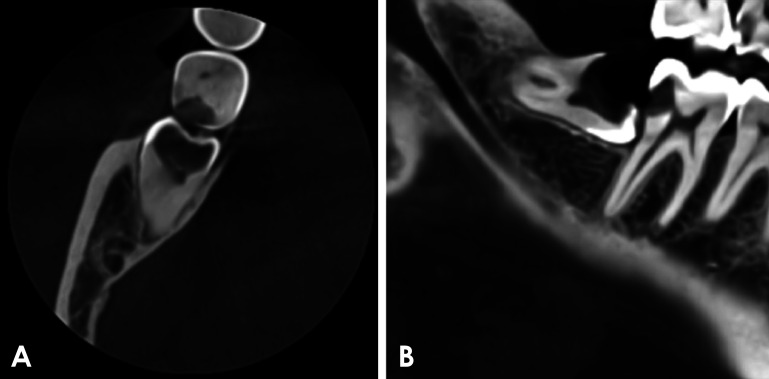
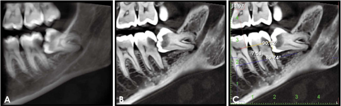
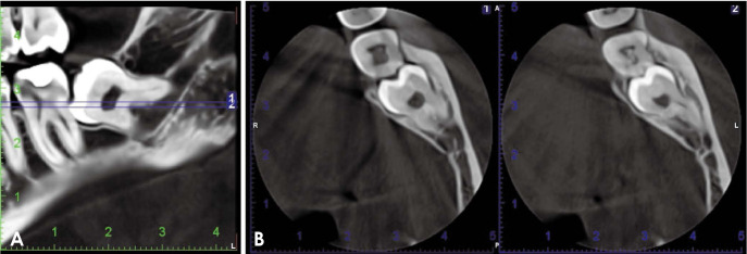
Materials and methods: Cone-beam computed tomographic images of impacted lower third molars were selected according to specific inclusion criteria. Impacted teeth were classified according to their position before assessment. The adjacent second molars were assessed for distal caries, distal bone loss, and root resorption. The fourth finding was the presence of a retromolar canal distal to the impaction. Communication with the dentist responsible for each case was done to determine whether these findings were detected or undetected by them before communication.
Results: Statistically significant correlations were found between impaction position, distal bone loss, and detected distal caries associated with the adjacent second molar. The greatest percentage of undetected findings was found in the evaluation of distal bone status, followed by missed detection of the retromolar canal.
Conclusion: The radiographic assessment protocol for impacted third molars should consider a step-by-step evaluation for second molars, and clinicians should be aware of the high prevalence of second molar affection in horizontal and mesioangular impactions. They also should search for the retromolar canal due to its associated clinical considerations.


 分享
分享
 求助内容:
求助内容: 应助结果提醒方式:
 扫码关注我们
