Yitong Yang, Zahraw Shah, Athira J Jacob, Jackson Hair, Teodora Chitiboi, Tiziano Passerini, Jerome Yerly, Lorenzo Di Sopra, Davide Piccini, Zahra Hosseini, Puneet Sharma, Anurag Sahu, Matthias Stuber, John N Oshinski
{"title":"Deep learning-based left ventricular segmentation demonstrates improved performance on respiratory motion-resolved whole-heart reconstructions.","authors":"Yitong Yang, Zahraw Shah, Athira J Jacob, Jackson Hair, Teodora Chitiboi, Tiziano Passerini, Jerome Yerly, Lorenzo Di Sopra, Davide Piccini, Zahra Hosseini, Puneet Sharma, Anurag Sahu, Matthias Stuber, John N Oshinski","doi":"10.3389/fradi.2023.1144004","DOIUrl":null,"url":null,"abstract":"<p><strong>Introduction: </strong>Deep learning (DL)-based segmentation has gained popularity for routine cardiac magnetic resonance (CMR) image analysis and in particular, delineation of left ventricular (LV) borders for LV volume determination. Free-breathing, self-navigated, whole-heart CMR exams provide high-resolution, isotropic coverage of the heart for assessment of cardiac anatomy including LV volume. The combination of whole-heart free-breathing CMR and DL-based LV segmentation has the potential to streamline the acquisition and analysis of clinical CMR exams. The purpose of this study was to compare the performance of a DL-based automatic LV segmentation network trained primarily on computed tomography (CT) images in two whole-heart CMR reconstruction methods: (1) an in-line respiratory motion-corrected (Mcorr) reconstruction and (2) an off-line, compressed sensing-based, multi-volume respiratory motion-resolved (Mres) reconstruction. Given that Mres images were shown to have greater image quality in previous studies than Mcorr images, we <i>hypothesized</i> that the LV volumes segmented from Mres images are closer to the manual expert-traced left ventricular endocardial border than the Mcorr images.</p><p><strong>Method: </strong>This retrospective study used 15 patients who underwent clinically indicated 1.5 T CMR exams with a prototype ECG-gated 3D radial phyllotaxis balanced steady state free precession (bSSFP) sequence. For each reconstruction method, the absolute volume difference (AVD) of the automatically and manually segmented LV volumes was used as the primary quantity to investigate whether 3D DL-based LV segmentation generalized better on Mcorr or Mres 3D whole-heart images. Additionally, we assessed the 3D Dice similarity coefficient between the manual and automatic LV masks of each reconstructed 3D whole-heart image and the sharpness of the LV myocardium-blood pool interface. A two-tail paired Student's <i>t</i>-test (alpha = 0.05) was used to test the significance in this study.</p><p><strong>Results & discussion: </strong>The AVD in the respiratory Mres reconstruction was lower than the AVD in the respiratory Mcorr reconstruction: 7.73 ± 6.54 ml vs. 20.0 ± 22.4 ml, respectively (<i>n</i> = 15, <i>p</i>-value = 0.03). The 3D Dice coefficient between the DL-segmented masks and the manually segmented masks was higher for Mres images than for Mcorr images: 0.90 ± 0.02 vs. 0.87 ± 0.03 respectively, with a <i>p</i>-value = 0.02. Sharpness on Mres images was higher than on Mcorr images: 0.15 ± 0.05 vs. 0.12 ± 0.04, respectively, with a <i>p</i>-value of 0.014 (<i>n</i> = 15).</p><p><strong>Conclusion: </strong>We conclude that the DL-based 3D automatic LV segmentation network trained on CT images and fine-tuned on MR images generalized better on Mres images than on Mcorr images for quantifying LV volumes.</p>","PeriodicalId":73101,"journal":{"name":"Frontiers in radiology","volume":"3 ","pages":"1144004"},"PeriodicalIF":2.3000,"publicationDate":"2023-06-02","publicationTypes":"Journal Article","fieldsOfStudy":null,"isOpenAccess":false,"openAccessPdf":"https://www.ncbi.nlm.nih.gov/pmc/articles/PMC10365088/pdf/","citationCount":"0","resultStr":null,"platform":"Semanticscholar","paperid":null,"PeriodicalName":"Frontiers in radiology","FirstCategoryId":"1085","ListUrlMain":"https://doi.org/10.3389/fradi.2023.1144004","RegionNum":0,"RegionCategory":null,"ArticlePicture":[],"TitleCN":null,"AbstractTextCN":null,"PMCID":null,"EPubDate":"2023/1/1 0:00:00","PubModel":"eCollection","JCR":"","JCRName":"","Score":null,"Total":0}
引用次数: 0
Abstract
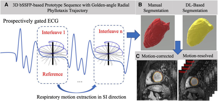
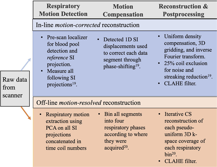
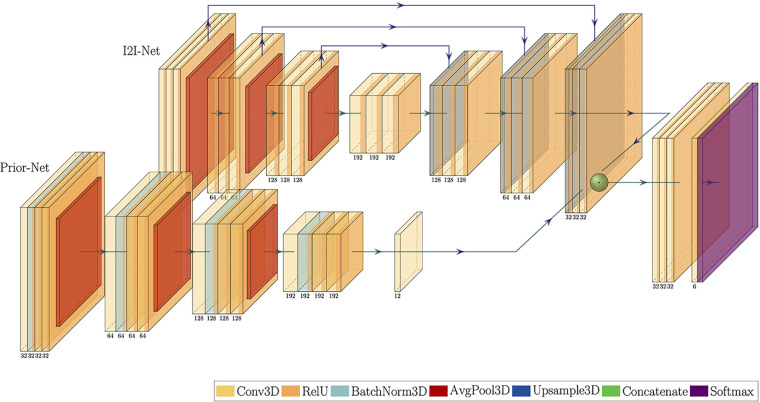
Introduction: Deep learning (DL)-based segmentation has gained popularity for routine cardiac magnetic resonance (CMR) image analysis and in particular, delineation of left ventricular (LV) borders for LV volume determination. Free-breathing, self-navigated, whole-heart CMR exams provide high-resolution, isotropic coverage of the heart for assessment of cardiac anatomy including LV volume. The combination of whole-heart free-breathing CMR and DL-based LV segmentation has the potential to streamline the acquisition and analysis of clinical CMR exams. The purpose of this study was to compare the performance of a DL-based automatic LV segmentation network trained primarily on computed tomography (CT) images in two whole-heart CMR reconstruction methods: (1) an in-line respiratory motion-corrected (Mcorr) reconstruction and (2) an off-line, compressed sensing-based, multi-volume respiratory motion-resolved (Mres) reconstruction. Given that Mres images were shown to have greater image quality in previous studies than Mcorr images, we hypothesized that the LV volumes segmented from Mres images are closer to the manual expert-traced left ventricular endocardial border than the Mcorr images.
Method: This retrospective study used 15 patients who underwent clinically indicated 1.5 T CMR exams with a prototype ECG-gated 3D radial phyllotaxis balanced steady state free precession (bSSFP) sequence. For each reconstruction method, the absolute volume difference (AVD) of the automatically and manually segmented LV volumes was used as the primary quantity to investigate whether 3D DL-based LV segmentation generalized better on Mcorr or Mres 3D whole-heart images. Additionally, we assessed the 3D Dice similarity coefficient between the manual and automatic LV masks of each reconstructed 3D whole-heart image and the sharpness of the LV myocardium-blood pool interface. A two-tail paired Student's t-test (alpha = 0.05) was used to test the significance in this study.
Results & discussion: The AVD in the respiratory Mres reconstruction was lower than the AVD in the respiratory Mcorr reconstruction: 7.73 ± 6.54 ml vs. 20.0 ± 22.4 ml, respectively (n = 15, p-value = 0.03). The 3D Dice coefficient between the DL-segmented masks and the manually segmented masks was higher for Mres images than for Mcorr images: 0.90 ± 0.02 vs. 0.87 ± 0.03 respectively, with a p-value = 0.02. Sharpness on Mres images was higher than on Mcorr images: 0.15 ± 0.05 vs. 0.12 ± 0.04, respectively, with a p-value of 0.014 (n = 15).
Conclusion: We conclude that the DL-based 3D automatic LV segmentation network trained on CT images and fine-tuned on MR images generalized better on Mres images than on Mcorr images for quantifying LV volumes.


 分享
分享

 求助内容:
求助内容: 应助结果提醒方式:
 扫码关注我们
