Ashlyn G Rickard, Yvonne M Mowery, Alex Bassil, Douglas C Rouse, Nerissa T Williams, Theresa Charity, Rafaela Belloni, Brian Crouch, Nimmi Ramanujam, Daniel Stevenson, Rico Castillo, Stephanie Blocker, Boris Epel, Mrignayani Kotecha, Gregory M Palmer
{"title":"Evaluating Tumor Hypoxia Radiosensitization Via Electron Paramagnetic Resonance Oxygen Imaging (EPROI).","authors":"Ashlyn G Rickard, Yvonne M Mowery, Alex Bassil, Douglas C Rouse, Nerissa T Williams, Theresa Charity, Rafaela Belloni, Brian Crouch, Nimmi Ramanujam, Daniel Stevenson, Rico Castillo, Stephanie Blocker, Boris Epel, Mrignayani Kotecha, Gregory M Palmer","doi":"10.1007/s11307-023-01855-0","DOIUrl":null,"url":null,"abstract":"<p><strong>Purpose: </strong>Tumor hypoxia contributes to aggressive phenotypes and diminished therapeutic responses to radiation therapy (RT) with hypoxic tissue being 3-fold less radiosensitive than normoxic tissue. A major challenge in implementing hypoxic radiosensitizers is the lack of a high-resolution imaging modality that directly quantifies tissue-oxygen. The electron paramagnetic resonance oxygen-imager (EPROI) was used to quantify tumor oxygenation in two murine tumor models: E0771 syngeneic transplant breast cancers and primary p53/MCA soft tissue sarcomas, with the latter autochthonous model better recapitulating the tumor microenvironment in human malignancies. We hypothesized that tumor hypoxia differs between these models. We also aimed to quantify the absolute change in tumor hypoxia induced by the mitochondrial inhibitor papaverine (PPV) and its effect on RT response.</p><p><strong>Procedures: </strong>Tumor oxygenation was characterized in E0771 and primary p53/MCA sarcomas via EPROI, with the former model also being quantified indirectly via diffuse reflectance spectroscopy (DRS). After confirming PPV's effect on hypoxic fraction (via EPROI), we compared the effect of 0 versus 2 mg/kg PPV prior to 20 Gy on tumor growth delay and survival.</p><p><strong>Results: </strong>Hypoxic sarcomas were more radioresistant than normoxic sarcomas (p=0.0057, 2-way ANOVA), and high baseline hypoxic fraction was a significant (p=0.0063, Cox Regression Model) hazard in survivability regardless of treatment. Pre-treatment with PPV before RT did not radiosensitize tumors in the sarcoma or E0771 model. In the sarcoma model, EPROI successfully identified baseline hypoxic tumors. DRS quantification of total hemoglobin, saturated hemoglobin, changes in mitochondrial potential and glucose uptake showed no significant difference in E0771 tumors pre- and post-PPV.</p><p><strong>Conclusion: </strong>EPROI provides 3D high-resolution pO<sub>2</sub> quantification; EPR is better suited than DRS to characterize tumor hypoxia. PPV did not radiosensitize E0771 tumors nor p53/MCA sarcomas, which may be related to the complex pattern of vasculature in each tumor. Additionally, understanding model-dependent tumor hypoxia will provide a much-needed foundation for future therapeutic studies with hypoxic radiosensitizers.</p>","PeriodicalId":18760,"journal":{"name":"Molecular Imaging and Biology","volume":" ","pages":"435-447"},"PeriodicalIF":2.5000,"publicationDate":"2024-06-01","publicationTypes":"Journal Article","fieldsOfStudy":null,"isOpenAccess":false,"openAccessPdf":"https://www.ncbi.nlm.nih.gov/pmc/articles/PMC12135767/pdf/","citationCount":"0","resultStr":null,"platform":"Semanticscholar","paperid":null,"PeriodicalName":"Molecular Imaging and Biology","FirstCategoryId":"3","ListUrlMain":"https://doi.org/10.1007/s11307-023-01855-0","RegionNum":4,"RegionCategory":"医学","ArticlePicture":[],"TitleCN":null,"AbstractTextCN":null,"PMCID":null,"EPubDate":"2023/9/18 0:00:00","PubModel":"Epub","JCR":"Q2","JCRName":"RADIOLOGY, NUCLEAR MEDICINE & MEDICAL IMAGING","Score":null,"Total":0}
引用次数: 0
Abstract
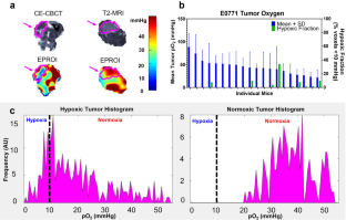
Purpose: Tumor hypoxia contributes to aggressive phenotypes and diminished therapeutic responses to radiation therapy (RT) with hypoxic tissue being 3-fold less radiosensitive than normoxic tissue. A major challenge in implementing hypoxic radiosensitizers is the lack of a high-resolution imaging modality that directly quantifies tissue-oxygen. The electron paramagnetic resonance oxygen-imager (EPROI) was used to quantify tumor oxygenation in two murine tumor models: E0771 syngeneic transplant breast cancers and primary p53/MCA soft tissue sarcomas, with the latter autochthonous model better recapitulating the tumor microenvironment in human malignancies. We hypothesized that tumor hypoxia differs between these models. We also aimed to quantify the absolute change in tumor hypoxia induced by the mitochondrial inhibitor papaverine (PPV) and its effect on RT response.
Procedures: Tumor oxygenation was characterized in E0771 and primary p53/MCA sarcomas via EPROI, with the former model also being quantified indirectly via diffuse reflectance spectroscopy (DRS). After confirming PPV's effect on hypoxic fraction (via EPROI), we compared the effect of 0 versus 2 mg/kg PPV prior to 20 Gy on tumor growth delay and survival.
Results: Hypoxic sarcomas were more radioresistant than normoxic sarcomas (p=0.0057, 2-way ANOVA), and high baseline hypoxic fraction was a significant (p=0.0063, Cox Regression Model) hazard in survivability regardless of treatment. Pre-treatment with PPV before RT did not radiosensitize tumors in the sarcoma or E0771 model. In the sarcoma model, EPROI successfully identified baseline hypoxic tumors. DRS quantification of total hemoglobin, saturated hemoglobin, changes in mitochondrial potential and glucose uptake showed no significant difference in E0771 tumors pre- and post-PPV.
Conclusion: EPROI provides 3D high-resolution pO2 quantification; EPR is better suited than DRS to characterize tumor hypoxia. PPV did not radiosensitize E0771 tumors nor p53/MCA sarcomas, which may be related to the complex pattern of vasculature in each tumor. Additionally, understanding model-dependent tumor hypoxia will provide a much-needed foundation for future therapeutic studies with hypoxic radiosensitizers.
期刊介绍:
Molecular Imaging and Biology (MIB) invites original contributions (research articles, review articles, commentaries, etc.) on the utilization of molecular imaging (i.e., nuclear imaging, optical imaging, autoradiography and pathology, MRI, MPI, ultrasound imaging, radiomics/genomics etc.) to investigate questions related to biology and health. The objective of MIB is to provide a forum to the discovery of molecular mechanisms of disease through the use of imaging techniques. We aim to investigate the biological nature of disease in patients and establish new molecular imaging diagnostic and therapy procedures.
Some areas that are covered are:
Preclinical and clinical imaging of macromolecular targets (e.g., genes, receptors, enzymes) involved in significant biological processes.
The design, characterization, and study of new molecular imaging probes and contrast agents for the functional interrogation of macromolecular targets.
Development and evaluation of imaging systems including instrumentation, image reconstruction algorithms, image analysis, and display.
Development of molecular assay approaches leading to quantification of the biological information obtained in molecular imaging.
Study of in vivo animal models of disease for the development of new molecular diagnostics and therapeutics.
Extension of in vitro and in vivo discoveries using disease models, into well designed clinical research investigations.
Clinical molecular imaging involving clinical investigations, clinical trials and medical management or cost-effectiveness studies.


 分享
分享

 求助内容:
求助内容: 应助结果提醒方式:
 扫码关注我们
