Nicholas M Jacobson, Erik Carerra, Aaron Treat, Megan McDonnell, David Mathes, Christodoulous Kaoutzanis
{"title":"Hybrid modeling techniques for 3D printed deep inferior epigastric perforator flap models.","authors":"Nicholas M Jacobson, Erik Carerra, Aaron Treat, Megan McDonnell, David Mathes, Christodoulous Kaoutzanis","doi":"10.1186/s41205-023-00181-z","DOIUrl":null,"url":null,"abstract":"<p><strong>Background: </strong>Deep Inferior Epigastric Perforator Flap (DIEP) surgical procedures have benefited in recent years from the introduction of 3D printed models, yet new technologies are expanding design opportunities which promise to improve patient specific care. Numerous studies, utilizing 3D printed models for DIEP, have shown a reduction of surgical time and complications when used in addition to the review of standard CT imaging. A DIEP free flap procedure requires locating the inferior epigastric perforator vessels traversing and perforating the rectus abdominis muscle, perfusing the abdominal skin and fatty tissue. The goal of dissecting the inferior epigastric perforator vessels is complicated by the opacity of the fatty tissue and muscle. Previous attempts to 3D print patient specific models for DIEP free flap cases from CT imaging has shown a wide range of designs which only show variations of perforator arteries, fatty tissue, and the abdominis rectus muscle.</p><p><strong>Methods: </strong>To remedy this limitation, we have leveraged a voxel-based modeling environment to composite complex modeling elements and incorporate a ruled grid upon the muscle providing effortless 'booleaning' and measured guidance.</p><p><strong>Results: </strong>A limitation of digital surface-based modeling tools has led to existing models lacking the ability to composite critical anatomical features, such as differentiation of vessels through different tissues, coherently into one model, providing information more akin to the surgical challenge.</p><p><strong>Conclusion: </strong>With new technology, highly detailed multi-material 3D printed models are allowing more of the information from medical imaging to be expressed in 3D printed models. This additional data, coupled with advanced digital modeling tools harnessing both voxel- and mesh-based modeling environments, is allowing for an expanded library of modeling techniques which create a wealth of concepts surgeons can use to assemble a presurgical planning model tailored to their setting, equipment, and needs.</p><p><strong>Trial registration: </strong>COMIRB 21-3135, ClinicalTrials.gov ID: NCT05144620.</p>","PeriodicalId":72036,"journal":{"name":"3D printing in medicine","volume":"9 1","pages":"26"},"PeriodicalIF":3.1000,"publicationDate":"2023-09-12","publicationTypes":"Journal Article","fieldsOfStudy":null,"isOpenAccess":false,"openAccessPdf":"https://www.ncbi.nlm.nih.gov/pmc/articles/PMC10498601/pdf/","citationCount":"0","resultStr":null,"platform":"Semanticscholar","paperid":null,"PeriodicalName":"3D printing in medicine","FirstCategoryId":"1085","ListUrlMain":"https://doi.org/10.1186/s41205-023-00181-z","RegionNum":0,"RegionCategory":null,"ArticlePicture":[],"TitleCN":null,"AbstractTextCN":null,"PMCID":null,"EPubDate":"","PubModel":"","JCR":"Q1","JCRName":"RADIOLOGY, NUCLEAR MEDICINE & MEDICAL IMAGING","Score":null,"Total":0}
引用次数: 0
Abstract
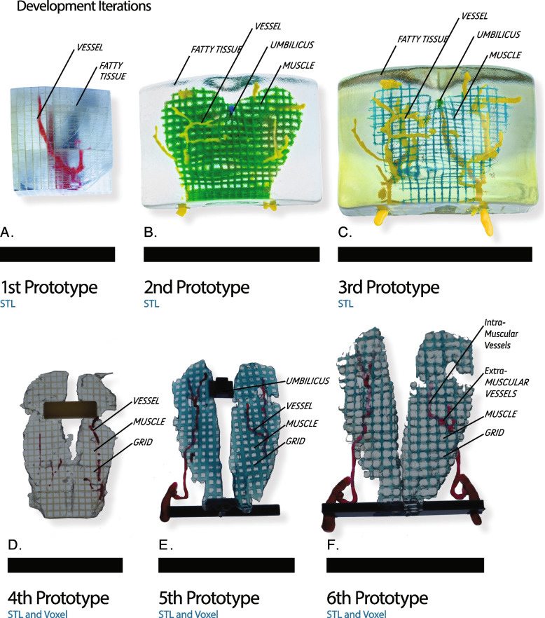
Background: Deep Inferior Epigastric Perforator Flap (DIEP) surgical procedures have benefited in recent years from the introduction of 3D printed models, yet new technologies are expanding design opportunities which promise to improve patient specific care. Numerous studies, utilizing 3D printed models for DIEP, have shown a reduction of surgical time and complications when used in addition to the review of standard CT imaging. A DIEP free flap procedure requires locating the inferior epigastric perforator vessels traversing and perforating the rectus abdominis muscle, perfusing the abdominal skin and fatty tissue. The goal of dissecting the inferior epigastric perforator vessels is complicated by the opacity of the fatty tissue and muscle. Previous attempts to 3D print patient specific models for DIEP free flap cases from CT imaging has shown a wide range of designs which only show variations of perforator arteries, fatty tissue, and the abdominis rectus muscle.
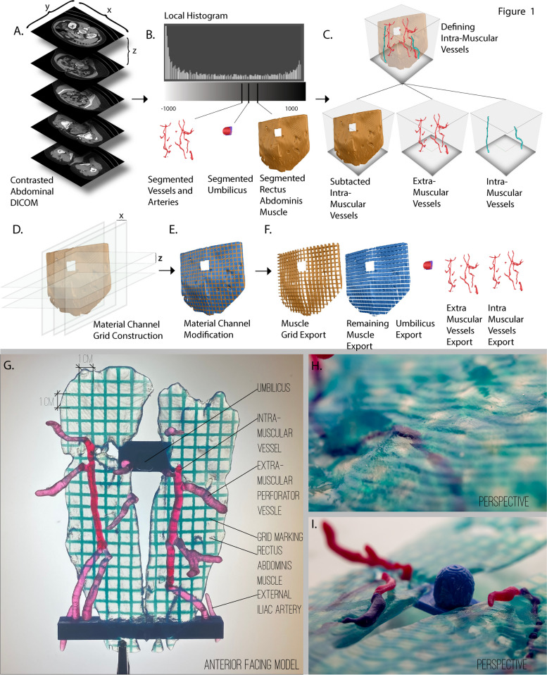
Methods: To remedy this limitation, we have leveraged a voxel-based modeling environment to composite complex modeling elements and incorporate a ruled grid upon the muscle providing effortless 'booleaning' and measured guidance.
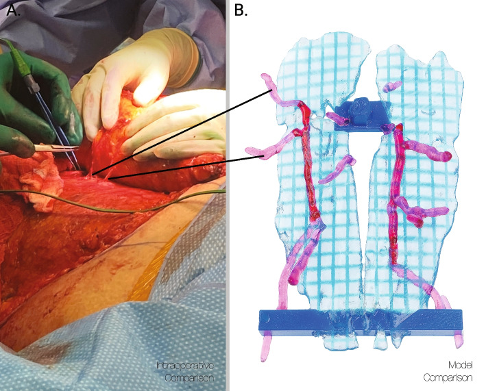
Results: A limitation of digital surface-based modeling tools has led to existing models lacking the ability to composite critical anatomical features, such as differentiation of vessels through different tissues, coherently into one model, providing information more akin to the surgical challenge.
Conclusion: With new technology, highly detailed multi-material 3D printed models are allowing more of the information from medical imaging to be expressed in 3D printed models. This additional data, coupled with advanced digital modeling tools harnessing both voxel- and mesh-based modeling environments, is allowing for an expanded library of modeling techniques which create a wealth of concepts surgeons can use to assemble a presurgical planning model tailored to their setting, equipment, and needs.


 分享
分享

 求助内容:
求助内容: 应助结果提醒方式:
 扫码关注我们
