{"title":"Low frequency 3D transmission ultrasound tomography: technical details and clinical implications","authors":"James Wiskin , Bilal Malik , John Klock","doi":"10.1016/j.zemedi.2023.04.006","DOIUrl":null,"url":null,"abstract":"<div><p>A novel 3D ultrasound tomographic (3D UT) method (called volography) that creates a speed of sound (SOS) map and a reflection modality that is co-registered are reviewed and shown to be artifact free even in the presence of high contrast and thus shown to be applicable for breast, orthopedic and pediatric clinical use cases. The 3D UT images are almost isotropic with mm resolution and the reflection image is compounded over 360 degrees to create sub-mm resolution in plane.</p></div><div><h3>Methods</h3><p>The physics of ultrasound scattering requires 3D modeling and the concomitant high computational cost is ameliorated with a bespoke algorithm (paraxial approximation – discussed here) and Nvidia GPUs. The resulting reconstruction times are tabulated for clinical relevance. The resulting SOS map is used to create a refraction corrected reflection image at ∼3.6 MHz center frequency. The transmission data are highly redundant, collected over 360 degrees and at 2 mm levels by true matrix receiver arrays yielding 3D data.</p><p>The high resolution SOS and attenuation maps and reflection images are used in a segmentation algorithm that optimally utilizes this information to segment out glandular, ductal, connective tissue, fat and skin. These volumes are used to estimate breast density, an important correlate to cancer.</p></div><div><h3>Results</h3><p>Multiple SOS images of breast, knee and segmentations of breast glandular and ductal tissue are shown. Spearman <em>rho</em> is calculated between our volumetric breast density estimates and Volpara™ from mammograms, as 0.9332. Multiple timing results are shown and indicate the variability of the reconstruction times with breast size and type but are ∼30 minutes for average size breast. The timing results with the 3D algorithm indicate ∼60 minute reconstruction times for pediatrics with two Nvidia GPUs. Characteristic variations of the glandular and ductal volumes over time are shown. The SOS from QT images are compared with literature values.</p><p>The results of a multi-reader multi-case (MRMC) study are shown that compares the 3D UT with full field digital mammography and resulted in an average increase in ROC AUC of 10%. Orthopedic (knee) 3D UT images compared with MRI indicate regions of zero signal in the MRI are clearly displayed in the QT image.</p><p>Explicit representation of the acoustic field is shown, indicating its 3D nature. An image of in vivo breast with the chest muscle is shown and speed of sound agreement with literature values are tabulated. Reference is made to a recently published paper validating pediatric imaging.</p></div><div><h3>Conclusions</h3><p>The high Spearman <em>rho</em> indicates a monotonic (not necessarily linear) relation between our method and industry gold standard Volpara™ density. The acoustic field verifies the need for 3D modeling. The MRMC study, the orthopedic images, breast density study, and references, all indicate the clinical utility of the SOS and reflection images. The QT image of the knee shows its ability to monitor tissue the MRI cannot. The included references and images herein indicate the proof of concept for 3D UT as a viable and valuable clinical adjunct in pediatric and orthopedic situations in addition to the breast imaging.</p></div>","PeriodicalId":54397,"journal":{"name":"Zeitschrift fur Medizinische Physik","volume":"33 3","pages":"Pages 427-443"},"PeriodicalIF":4.2000,"publicationDate":"2023-08-01","publicationTypes":"Journal Article","fieldsOfStudy":null,"isOpenAccess":false,"openAccessPdf":"https://ftp.ncbi.nlm.nih.gov/pub/pmc/oa_pdf/90/c2/main.PMC10517404.pdf","citationCount":"0","resultStr":null,"platform":"Semanticscholar","paperid":null,"PeriodicalName":"Zeitschrift fur Medizinische Physik","FirstCategoryId":"3","ListUrlMain":"https://www.sciencedirect.com/science/article/pii/S093938892300048X","RegionNum":4,"RegionCategory":"医学","ArticlePicture":[],"TitleCN":null,"AbstractTextCN":null,"PMCID":null,"EPubDate":"2023/6/7 0:00:00","PubModel":"Epub","JCR":"Q2","JCRName":"RADIOLOGY, NUCLEAR MEDICINE & MEDICAL IMAGING","Score":null,"Total":0}
引用次数: 0
Abstract
A novel 3D ultrasound tomographic (3D UT) method (called volography) that creates a speed of sound (SOS) map and a reflection modality that is co-registered are reviewed and shown to be artifact free even in the presence of high contrast and thus shown to be applicable for breast, orthopedic and pediatric clinical use cases. The 3D UT images are almost isotropic with mm resolution and the reflection image is compounded over 360 degrees to create sub-mm resolution in plane.
Methods
The physics of ultrasound scattering requires 3D modeling and the concomitant high computational cost is ameliorated with a bespoke algorithm (paraxial approximation – discussed here) and Nvidia GPUs. The resulting reconstruction times are tabulated for clinical relevance. The resulting SOS map is used to create a refraction corrected reflection image at ∼3.6 MHz center frequency. The transmission data are highly redundant, collected over 360 degrees and at 2 mm levels by true matrix receiver arrays yielding 3D data.
The high resolution SOS and attenuation maps and reflection images are used in a segmentation algorithm that optimally utilizes this information to segment out glandular, ductal, connective tissue, fat and skin. These volumes are used to estimate breast density, an important correlate to cancer.
Results
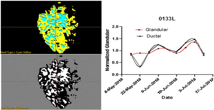
Multiple SOS images of breast, knee and segmentations of breast glandular and ductal tissue are shown. Spearman rho is calculated between our volumetric breast density estimates and Volpara™ from mammograms, as 0.9332. Multiple timing results are shown and indicate the variability of the reconstruction times with breast size and type but are ∼30 minutes for average size breast. The timing results with the 3D algorithm indicate ∼60 minute reconstruction times for pediatrics with two Nvidia GPUs. Characteristic variations of the glandular and ductal volumes over time are shown. The SOS from QT images are compared with literature values.
The results of a multi-reader multi-case (MRMC) study are shown that compares the 3D UT with full field digital mammography and resulted in an average increase in ROC AUC of 10%. Orthopedic (knee) 3D UT images compared with MRI indicate regions of zero signal in the MRI are clearly displayed in the QT image.
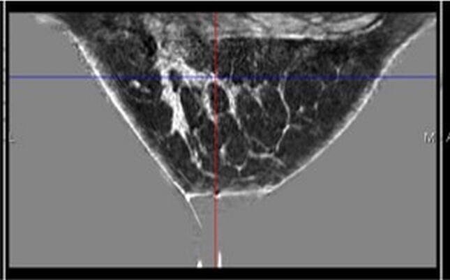
Explicit representation of the acoustic field is shown, indicating its 3D nature. An image of in vivo breast with the chest muscle is shown and speed of sound agreement with literature values are tabulated. Reference is made to a recently published paper validating pediatric imaging.
Conclusions
The high Spearman rho indicates a monotonic (not necessarily linear) relation between our method and industry gold standard Volpara™ density. The acoustic field verifies the need for 3D modeling. The MRMC study, the orthopedic images, breast density study, and references, all indicate the clinical utility of the SOS and reflection images. The QT image of the knee shows its ability to monitor tissue the MRI cannot. The included references and images herein indicate the proof of concept for 3D UT as a viable and valuable clinical adjunct in pediatric and orthopedic situations in addition to the breast imaging.
期刊介绍:
Zeitschrift fur Medizinische Physik (Journal of Medical Physics) is an official organ of the German and Austrian Society of Medical Physic and the Swiss Society of Radiobiology and Medical Physics.The Journal is a platform for basic research and practical applications of physical procedures in medical diagnostics and therapy. The articles are reviewed following international standards of peer reviewing.
Focuses of the articles are:
-Biophysical methods in radiation therapy and nuclear medicine
-Dosimetry and radiation protection
-Radiological diagnostics and quality assurance
-Modern imaging techniques, such as computed tomography, magnetic resonance imaging, positron emission tomography
-Ultrasonography diagnostics, application of laser and UV rays
-Electronic processing of biosignals
-Artificial intelligence and machine learning in medical physics
In the Journal, the latest scientific insights find their expression in the form of original articles, reviews, technical communications, and information for the clinical practice.


 分享
分享

 求助内容:
求助内容: 应助结果提醒方式:
 扫码关注我们
