{"title":"Ultrasonographic Characteristics in the Fingers and Other Superficial Glomus Tumours.","authors":"Noboru Takanashi, Satomi Asai, Yoko Ogase, Akiko Fujii, Haruyo Atsumi, Mika Doi, Nobue Kumaki, Tomotaka Mabuchi, Hayato Miyachi","doi":"10.1155/2023/7126799","DOIUrl":null,"url":null,"abstract":"<p><p>Glomus tumours are painful superficial tumours, and ultrasonography is an extremely useful and noninvasive diagnostic technique for superficial organs. In this study, we retrospectively examined glomus tumours using ultrasonography. Among 18 patients histopathologically diagnosed with glomus tumours via ultrasonography, we observed five different development sites: subungual areas or those surrounding the nail bed (12), other areas on the finger surface (3), abdominal wall (1), upper arm (1), and forearm (1). The ultrasonographic images revealed significant differences in tumour size, indicating that tumours on other body surfaces tended to be smaller than those on patients' fingers (<i>p</i> < 0.01). The depth/width ratios of tumours on the other body surfaces were significantly higher than those on the fingers (<i>p</i> < 0.05). The tumours showed a regular shape (72.2%) and clear border (100%). Furthermore, most tumours were low-echo tumours with a diameter of up to 15 mm, clear margins, and no lateral shadows. Abundant blood flow and vessels in and out of the tumours were also observed. In conclusion, our study describes the ultrasonographic characteristics of glomus tumours and reveals that they cannot be ruled out when diagnosing small painful subcutaneous tumours.</p>","PeriodicalId":11338,"journal":{"name":"Dermatology Research and Practice","volume":"2023 ","pages":"7126799"},"PeriodicalIF":1.9000,"publicationDate":"2023-01-01","publicationTypes":"Journal Article","fieldsOfStudy":null,"isOpenAccess":false,"openAccessPdf":"https://www.ncbi.nlm.nih.gov/pmc/articles/PMC10397493/pdf/","citationCount":"0","resultStr":null,"platform":"Semanticscholar","paperid":null,"PeriodicalName":"Dermatology Research and Practice","FirstCategoryId":"1085","ListUrlMain":"https://doi.org/10.1155/2023/7126799","RegionNum":0,"RegionCategory":null,"ArticlePicture":[],"TitleCN":null,"AbstractTextCN":null,"PMCID":null,"EPubDate":"","PubModel":"","JCR":"Q3","JCRName":"DERMATOLOGY","Score":null,"Total":0}
引用次数: 0
Abstract
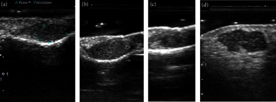
Glomus tumours are painful superficial tumours, and ultrasonography is an extremely useful and noninvasive diagnostic technique for superficial organs. In this study, we retrospectively examined glomus tumours using ultrasonography. Among 18 patients histopathologically diagnosed with glomus tumours via ultrasonography, we observed five different development sites: subungual areas or those surrounding the nail bed (12), other areas on the finger surface (3), abdominal wall (1), upper arm (1), and forearm (1). The ultrasonographic images revealed significant differences in tumour size, indicating that tumours on other body surfaces tended to be smaller than those on patients' fingers (p < 0.01). The depth/width ratios of tumours on the other body surfaces were significantly higher than those on the fingers (p < 0.05). The tumours showed a regular shape (72.2%) and clear border (100%). Furthermore, most tumours were low-echo tumours with a diameter of up to 15 mm, clear margins, and no lateral shadows. Abundant blood flow and vessels in and out of the tumours were also observed. In conclusion, our study describes the ultrasonographic characteristics of glomus tumours and reveals that they cannot be ruled out when diagnosing small painful subcutaneous tumours.


 分享
分享
 求助内容:
求助内容: 应助结果提醒方式:
 扫码关注我们
