Boris Epel, Navin Viswakarma, Subramanian V Sundramoorthy, Nitin J Pawar, Mrignayani Kotecha
{"title":"Oxygen Imaging of a Rabbit Tumor Using a Human-Sized Pulse Electron Paramagnetic Resonance Imager.","authors":"Boris Epel, Navin Viswakarma, Subramanian V Sundramoorthy, Nitin J Pawar, Mrignayani Kotecha","doi":"10.1007/s11307-023-01852-3","DOIUrl":null,"url":null,"abstract":"<p><strong>Purpose: </strong>Spatial heterogeneity in tumor hypoxia is one of the most important factors regulating tumor growth, development, aggressiveness, metastasis, and affecting treatment outcome. Most solid tumors are known to have hypoxia or low oxygen levels (pO<sub>2</sub> ≤10 torr). Electron paramagnetic resonance oxygen imaging (EPROI) is an emerging oxygen mapping technology. EPROI utilizes the linear relationship between the relaxation rates of the injectable OX071 trityl spin probe and the partial oxygen pressure (pO<sub>2</sub>). However, most of the EPROI studies have been limited to mouse models of solid tumors because of the instrument-size limitations. The purpose of this work was to develop a human-sized 9-mT (250 MHz resonance frequency, 60 cm bore size) pulse EPROI instrument and evaluate its performance with rabbit VX-2 tumor oxygen imaging.</p><p><strong>Methods: </strong>A New Zealand white rabbit with a 3.2-cm VX-2 tumor in the calf muscle was imaged using the human-sized EPROI instrument and a 2.25-in. ID volume coil. The animal received a ~8-min intravenous injection of OX071 (5.2 mL total volume at 72 mM concentration) and, after 75 min, an intratumoral injection (120 μL total at 5 mM OX071 concentration) and underwent EPROI. At the end of the experiments, MRI was performed using a preclinical 9.4-T MRI system to outline the tumor boundaries.</p><p><strong>Results: </strong>For the first time, a human-sized pulse EPROI instrument with a 60-cm bore size/250-MHz frequency was built and evaluated using rabbit tumor oxygen imaging. For the first time, the systemic IV injection of the oxygen-sensitive trityl OX071 spin probe was used for an animal of this size. The resulting EPROI image from the IV injection showed complete tumor coverage. The image obtained after intratumoral injection showed localized coverage in the upper lobe of the tumor, demonstrating the need for improved intratumoral injection protocol.</p><p><strong>Conclusions: </strong>This study demonstrates the performance of the world's first human-sized pulse EPROI instrument. It also demonstrates that the EPROI of larger animals can be performed using the systemic injection of a manageable amount of the spin probe. This brings EPROI one step closer to clinical applications in cancer therapies. Oxygen imaging is a platform technology, and the instrument and techniques developed here will also be useful for other clinical applications.</p>","PeriodicalId":18760,"journal":{"name":"Molecular Imaging and Biology","volume":" ","pages":"403-410"},"PeriodicalIF":2.5000,"publicationDate":"2024-06-01","publicationTypes":"Journal Article","fieldsOfStudy":null,"isOpenAccess":false,"openAccessPdf":"","citationCount":"0","resultStr":null,"platform":"Semanticscholar","paperid":null,"PeriodicalName":"Molecular Imaging and Biology","FirstCategoryId":"3","ListUrlMain":"https://doi.org/10.1007/s11307-023-01852-3","RegionNum":4,"RegionCategory":"医学","ArticlePicture":[],"TitleCN":null,"AbstractTextCN":null,"PMCID":null,"EPubDate":"2023/9/15 0:00:00","PubModel":"Epub","JCR":"Q2","JCRName":"RADIOLOGY, NUCLEAR MEDICINE & MEDICAL IMAGING","Score":null,"Total":0}
引用次数: 0
Abstract
Purpose: Spatial heterogeneity in tumor hypoxia is one of the most important factors regulating tumor growth, development, aggressiveness, metastasis, and affecting treatment outcome. Most solid tumors are known to have hypoxia or low oxygen levels (pO2 ≤10 torr). Electron paramagnetic resonance oxygen imaging (EPROI) is an emerging oxygen mapping technology. EPROI utilizes the linear relationship between the relaxation rates of the injectable OX071 trityl spin probe and the partial oxygen pressure (pO2). However, most of the EPROI studies have been limited to mouse models of solid tumors because of the instrument-size limitations. The purpose of this work was to develop a human-sized 9-mT (250 MHz resonance frequency, 60 cm bore size) pulse EPROI instrument and evaluate its performance with rabbit VX-2 tumor oxygen imaging.
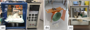
Methods: A New Zealand white rabbit with a 3.2-cm VX-2 tumor in the calf muscle was imaged using the human-sized EPROI instrument and a 2.25-in. ID volume coil. The animal received a ~8-min intravenous injection of OX071 (5.2 mL total volume at 72 mM concentration) and, after 75 min, an intratumoral injection (120 μL total at 5 mM OX071 concentration) and underwent EPROI. At the end of the experiments, MRI was performed using a preclinical 9.4-T MRI system to outline the tumor boundaries.
Results: For the first time, a human-sized pulse EPROI instrument with a 60-cm bore size/250-MHz frequency was built and evaluated using rabbit tumor oxygen imaging. For the first time, the systemic IV injection of the oxygen-sensitive trityl OX071 spin probe was used for an animal of this size. The resulting EPROI image from the IV injection showed complete tumor coverage. The image obtained after intratumoral injection showed localized coverage in the upper lobe of the tumor, demonstrating the need for improved intratumoral injection protocol.
Conclusions: This study demonstrates the performance of the world's first human-sized pulse EPROI instrument. It also demonstrates that the EPROI of larger animals can be performed using the systemic injection of a manageable amount of the spin probe. This brings EPROI one step closer to clinical applications in cancer therapies. Oxygen imaging is a platform technology, and the instrument and techniques developed here will also be useful for other clinical applications.
期刊介绍:
Molecular Imaging and Biology (MIB) invites original contributions (research articles, review articles, commentaries, etc.) on the utilization of molecular imaging (i.e., nuclear imaging, optical imaging, autoradiography and pathology, MRI, MPI, ultrasound imaging, radiomics/genomics etc.) to investigate questions related to biology and health. The objective of MIB is to provide a forum to the discovery of molecular mechanisms of disease through the use of imaging techniques. We aim to investigate the biological nature of disease in patients and establish new molecular imaging diagnostic and therapy procedures.
Some areas that are covered are:
Preclinical and clinical imaging of macromolecular targets (e.g., genes, receptors, enzymes) involved in significant biological processes.
The design, characterization, and study of new molecular imaging probes and contrast agents for the functional interrogation of macromolecular targets.
Development and evaluation of imaging systems including instrumentation, image reconstruction algorithms, image analysis, and display.
Development of molecular assay approaches leading to quantification of the biological information obtained in molecular imaging.
Study of in vivo animal models of disease for the development of new molecular diagnostics and therapeutics.
Extension of in vitro and in vivo discoveries using disease models, into well designed clinical research investigations.
Clinical molecular imaging involving clinical investigations, clinical trials and medical management or cost-effectiveness studies.


 分享
分享

 求助内容:
求助内容: 应助结果提醒方式:
 扫码关注我们
