Magdalena Chylińska, Bartosz Karaszewski, Jakub Komendziński, Adam Wyszomirski, Marek Hałas, Edyta Szurowska, Agnieszka Sabisz
{"title":"The association between white matter tract structural connectivity and information processing speed in relapsing-remitting multiple sclerosis.","authors":"Magdalena Chylińska, Bartosz Karaszewski, Jakub Komendziński, Adam Wyszomirski, Marek Hałas, Edyta Szurowska, Agnieszka Sabisz","doi":"10.1007/s10072-023-06817-6","DOIUrl":null,"url":null,"abstract":"<p><strong>Background: </strong>Information processing speed (IPS) deterioration is common in relapsing-remitting multiple sclerosis (RRMS) patients [1] and might severely affect quality of life and occupational activity. However, understanding of its neural substrate is not fully elucidated. We aimed to investigate the associations between MRI-derived metrics of neuroanatomical structures, including the tracts, and IPS.</p><p><strong>Methods: </strong>Symbol Digit Modalities Test (SDMT), Paced Auditory Serial Addition Test (PASAT), and Color Trails Test (CTT) were used to evaluate IPS in 73 RRMS consecutive patients, all undergoing only interferon beta (IFN-β) therapy during the study. At the same time, 1.5T MRI including diffusion tensor imaging (DTI) data was acquired for each recruited subject. We analyzed volumetric and diffusion MRI measures (FreeSurfer 6.0) including normalized brain volume (NBV), cortical thickness (thk), white matter hypointensities (WMH), volume (vol), diffusion parameters: mean (MD), radial (RD), axial (AD) diffusivities, and fractional anisotropy (FA) of 18 major white-matter (WM) tracts. Multiple linear regression model with interaction resulted in distinguishing the neural substrate of IPS deficit in the IPS impaired subgroup of patients.</p><p><strong>Results: </strong>The most significant tract abnormalities contributing to IPS deficit were right inferior longitudinal fasciculus (R ILF) FA, forceps major (FMAJ) FA, forceps minor (FMIN) FA, R uncinate fasciculus (UNC) AD, R corticospinal tract (CST) FA, and left superior longitudinal fasciculus FA (L SLFT). Among volumetric MRI metrics, IPS deficit was associated with L and R thalamic vol. and cortical thickness of insular regions.</p><p><strong>Conclusion: </strong>In this study, we showed that disconnection of the selected WM tracts, in addition to cortical and deep gray matter (GM) atrophy, might underlie IPS deficit in RRMS patients but more extensive studies are needed for precise associations.</p>","PeriodicalId":19191,"journal":{"name":"Neurological Sciences","volume":"44 9","pages":"3221-3232"},"PeriodicalIF":2.4000,"publicationDate":"2023-09-01","publicationTypes":"Journal Article","fieldsOfStudy":null,"isOpenAccess":false,"openAccessPdf":"https://www.ncbi.nlm.nih.gov/pmc/articles/PMC10415523/pdf/","citationCount":"0","resultStr":null,"platform":"Semanticscholar","paperid":null,"PeriodicalName":"Neurological Sciences","FirstCategoryId":"3","ListUrlMain":"https://doi.org/10.1007/s10072-023-06817-6","RegionNum":4,"RegionCategory":"医学","ArticlePicture":[],"TitleCN":null,"AbstractTextCN":null,"PMCID":null,"EPubDate":"2023/4/27 0:00:00","PubModel":"Epub","JCR":"Q2","JCRName":"CLINICAL NEUROLOGY","Score":null,"Total":0}
引用次数: 0
Abstract
Background: Information processing speed (IPS) deterioration is common in relapsing-remitting multiple sclerosis (RRMS) patients [1] and might severely affect quality of life and occupational activity. However, understanding of its neural substrate is not fully elucidated. We aimed to investigate the associations between MRI-derived metrics of neuroanatomical structures, including the tracts, and IPS.
Methods: Symbol Digit Modalities Test (SDMT), Paced Auditory Serial Addition Test (PASAT), and Color Trails Test (CTT) were used to evaluate IPS in 73 RRMS consecutive patients, all undergoing only interferon beta (IFN-β) therapy during the study. At the same time, 1.5T MRI including diffusion tensor imaging (DTI) data was acquired for each recruited subject. We analyzed volumetric and diffusion MRI measures (FreeSurfer 6.0) including normalized brain volume (NBV), cortical thickness (thk), white matter hypointensities (WMH), volume (vol), diffusion parameters: mean (MD), radial (RD), axial (AD) diffusivities, and fractional anisotropy (FA) of 18 major white-matter (WM) tracts. Multiple linear regression model with interaction resulted in distinguishing the neural substrate of IPS deficit in the IPS impaired subgroup of patients.
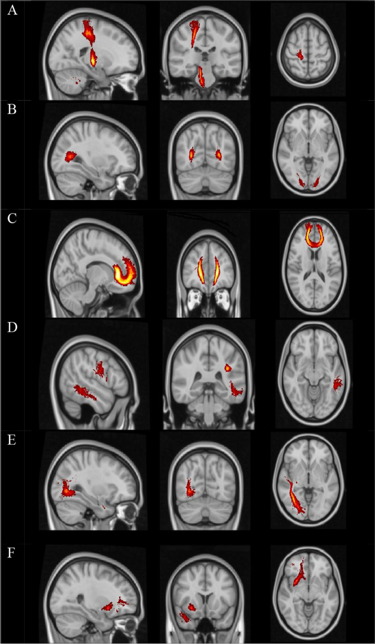
Results: The most significant tract abnormalities contributing to IPS deficit were right inferior longitudinal fasciculus (R ILF) FA, forceps major (FMAJ) FA, forceps minor (FMIN) FA, R uncinate fasciculus (UNC) AD, R corticospinal tract (CST) FA, and left superior longitudinal fasciculus FA (L SLFT). Among volumetric MRI metrics, IPS deficit was associated with L and R thalamic vol. and cortical thickness of insular regions.
Conclusion: In this study, we showed that disconnection of the selected WM tracts, in addition to cortical and deep gray matter (GM) atrophy, might underlie IPS deficit in RRMS patients but more extensive studies are needed for precise associations.
期刊介绍:
Neurological Sciences is intended to provide a medium for the communication of results and ideas in the field of neuroscience. The journal welcomes contributions in both the basic and clinical aspects of the neurosciences. The official language of the journal is English. Reports are published in the form of original articles, short communications, editorials, reviews and letters to the editor. Original articles present the results of experimental or clinical studies in the neurosciences, while short communications are succinct reports permitting the rapid publication of novel results. Original contributions may be submitted for the special sections History of Neurology, Health Care and Neurological Digressions - a forum for cultural topics related to the neurosciences. The journal also publishes correspondence book reviews, meeting reports and announcements.


 分享
分享
 求助内容:
求助内容: 应助结果提醒方式:
 扫码关注我们
