{"title":"Defining the Specialized Functions of cAMP Signals in an Organelle Formerly Deemed to Have No Function: The Primary Cilium.","authors":"Aldebaran M Hofer","doi":"10.1093/function/zqad007","DOIUrl":null,"url":null,"abstract":"The primary cilium is a decidedly “cute” organelle that first attracted the attention of electron microscopists in the early 1960s (Figure 1). A few prescient scientists of the last century recognized that the cilium was more than just an ultrastructural curiosity. But this was not the prevailing view; in fact, cell biology textbooks published only three decades ago discounted the cilium as a “vestigial” structure of “unknown function.” Interest in this diminutive organelle began to seriously build around 20 yr ago with the discovery of its central role in several disease states. These included autosomal dominant polycystic kidney disease (ADPKD), and inherited “ciliopathies” such as Bardet– Biedl syndrome. The ciliary localization of signaling pathways such as hedgehog and its connection to glioblastoma accelerated further interest.1 Now, in 2023, the primary cilium has been launched from relative obscurity to the focus of exuberant scientific inquiry. Discoveries of new connections between ciliary biology and cellular functions (autophagy, neuronal migration, and necroptosis, to name a few) seem to emerge daily. Loss of proper function is implicated in an ever-expanding list of human diseases and conditions that include obesity and appetite control, cancer, cognitive decline, and diabetes.1 Among the most titillating of observations to come to light in the past years (for some of us, at least), was that cilia are enriched with a specific subset of G-protein coupled receptors (GPCRs) that are typically linked to cAMP signaling through Galpha(s) or Galpha(i).2 Another early observation regarded the very exclusive sequestration of adenylyl cyclase 3 (AC3) in neuronal cilia, and its link to obesity and depression. Other AC isoforms (eg, AC5 and AC6) are now known to localize to cilia in different cell types, along with phosphodiesterases and betaarrestins. The main effector of the cAMP signal, protein kinase A (PKA), is tethered within the organelle by A-kinase anchoring proteins (AKAPs), ready to phosphorylate a host of potential and confirmed ciliary PKA targets. Direct measurements by our lab using targeted cAMP biosensors showed that stimulation of GPCRs confined exclusively in the cilium produced local cAMP signals. (Interestingly, this response was greatly amplified during activation of the hedgehog pathway.)3 Taken together, it appears everything is in place to operate a self-contained cAMP signaling circuit in the cilium. But what special purpose does it serve, especially considering that small molecules like cyclic nucleotides move freely between cytosol and cilium? Recent studies now implicate ciliary GPCRs coupled to cAMP production in the control of several physiological functions. For example, Hilgendorf et al. reported that white adipose tissue expansion and differentiation required FFAR4, a ciliary GPCR that could be stimulated by omega-3 fatty acids to locally produce cAMP.4 FFAR4 stimulation was shown to converge on the chromatin remodeling protein, CTFC, to turn on the adipogenic transcriptional program. Since FFAR4 was localized only to cilia, the presumption was that these effects were initiated by the ciliary cAMP signal.","PeriodicalId":73119,"journal":{"name":"Function (Oxford, England)","volume":"4 2","pages":"zqad007"},"PeriodicalIF":3.8000,"publicationDate":"2023-01-01","publicationTypes":"Journal Article","fieldsOfStudy":null,"isOpenAccess":false,"openAccessPdf":"https://ftp.ncbi.nlm.nih.gov/pub/pmc/oa_pdf/89/d4/zqad007.PMC9972343.pdf","citationCount":"0","resultStr":null,"platform":"Semanticscholar","paperid":null,"PeriodicalName":"Function (Oxford, England)","FirstCategoryId":"1085","ListUrlMain":"https://doi.org/10.1093/function/zqad007","RegionNum":0,"RegionCategory":null,"ArticlePicture":[],"TitleCN":null,"AbstractTextCN":null,"PMCID":null,"EPubDate":"","PubModel":"","JCR":"Q2","JCRName":"CELL BIOLOGY","Score":null,"Total":0}
引用次数: 0
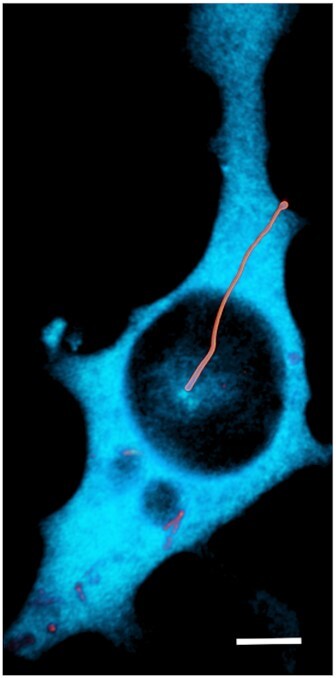
Abstract
The primary cilium is a decidedly “cute” organelle that first attracted the attention of electron microscopists in the early 1960s (Figure 1). A few prescient scientists of the last century recognized that the cilium was more than just an ultrastructural curiosity. But this was not the prevailing view; in fact, cell biology textbooks published only three decades ago discounted the cilium as a “vestigial” structure of “unknown function.” Interest in this diminutive organelle began to seriously build around 20 yr ago with the discovery of its central role in several disease states. These included autosomal dominant polycystic kidney disease (ADPKD), and inherited “ciliopathies” such as Bardet– Biedl syndrome. The ciliary localization of signaling pathways such as hedgehog and its connection to glioblastoma accelerated further interest.1 Now, in 2023, the primary cilium has been launched from relative obscurity to the focus of exuberant scientific inquiry. Discoveries of new connections between ciliary biology and cellular functions (autophagy, neuronal migration, and necroptosis, to name a few) seem to emerge daily. Loss of proper function is implicated in an ever-expanding list of human diseases and conditions that include obesity and appetite control, cancer, cognitive decline, and diabetes.1 Among the most titillating of observations to come to light in the past years (for some of us, at least), was that cilia are enriched with a specific subset of G-protein coupled receptors (GPCRs) that are typically linked to cAMP signaling through Galpha(s) or Galpha(i).2 Another early observation regarded the very exclusive sequestration of adenylyl cyclase 3 (AC3) in neuronal cilia, and its link to obesity and depression. Other AC isoforms (eg, AC5 and AC6) are now known to localize to cilia in different cell types, along with phosphodiesterases and betaarrestins. The main effector of the cAMP signal, protein kinase A (PKA), is tethered within the organelle by A-kinase anchoring proteins (AKAPs), ready to phosphorylate a host of potential and confirmed ciliary PKA targets. Direct measurements by our lab using targeted cAMP biosensors showed that stimulation of GPCRs confined exclusively in the cilium produced local cAMP signals. (Interestingly, this response was greatly amplified during activation of the hedgehog pathway.)3 Taken together, it appears everything is in place to operate a self-contained cAMP signaling circuit in the cilium. But what special purpose does it serve, especially considering that small molecules like cyclic nucleotides move freely between cytosol and cilium? Recent studies now implicate ciliary GPCRs coupled to cAMP production in the control of several physiological functions. For example, Hilgendorf et al. reported that white adipose tissue expansion and differentiation required FFAR4, a ciliary GPCR that could be stimulated by omega-3 fatty acids to locally produce cAMP.4 FFAR4 stimulation was shown to converge on the chromatin remodeling protein, CTFC, to turn on the adipogenic transcriptional program. Since FFAR4 was localized only to cilia, the presumption was that these effects were initiated by the ciliary cAMP signal.


 分享
分享

 求助内容:
求助内容: 应助结果提醒方式:
 扫码关注我们
