Lorenzo Marcucci, Antonio Michelucci, Carlo Reggiani
{"title":"Cytosolic Ca<sup>2+</sup> gradients and mitochondrial Ca<sup>2+</sup> uptake in resting muscle fibers: A model analysis.","authors":"Lorenzo Marcucci, Antonio Michelucci, Carlo Reggiani","doi":"10.1016/j.bpr.2023.100117","DOIUrl":null,"url":null,"abstract":"<p><p>Calcium ions (Ca<sup>2+</sup>) enter mitochondria via the mitochondrial Ca<sup>2+</sup> uniporter, driven by electrical and concentration gradients. In this regard, transgenic mouse models, such as calsequestrin knockout (CSQ-KO) mice, with higher mitochondrial Ca<sup>2+</sup> concentrations ([Ca<sup>2+</sup>]<sub>mito</sub>), should display higher cytosolic Ca<sup>2+</sup> concentrations ([Ca<sup>2+</sup>]<sub>cyto</sub>). However, repeated measurements of [Ca<sup>2+</sup>]<sub>cyto</sub> in quiescent CSQ-KO fibers never showed a difference between WT and CSQ-KO. Starting from the consideration that fluorescent Ca<sup>2+</sup> probes (Fura-2 and Indo-1) measure averaged global cytosolic concentrations, in this report we explored the role of local Ca<sup>2+</sup> concentrations (i.e., Ca<sup>2+</sup> microdomains) in regulating mitochondrial Ca<sup>2+</sup> in resting cells, using a multicompartmental diffusional Ca<sup>2+</sup> model. Progressively including the inward and outward fluxes of sarcoplasmic reticulum (SR), extracellular space, and mitochondria, we explored their contribution to the local Ca<sup>2+</sup> distribution within the cell. The model predicts Ca<sup>2+</sup> concentration gradients with hot spots or microdomains even at rest, minor but similar to those of evoked Ca<sup>2+</sup> release. Due to their specific localization close to Ca<sup>2+</sup> release units (CRU), mitochondria could take up Ca<sup>2+</sup> directly from high-concentration microdomains, thus sensibly raising [Ca<sup>2+</sup>]<sub>mito</sub>, despite minor, possibly undetectable, modifications of the average [Ca<sup>2+</sup>]<sub>cyto</sub>.</p>","PeriodicalId":72402,"journal":{"name":"Biophysical reports","volume":"3 3","pages":"100117"},"PeriodicalIF":2.7000,"publicationDate":"2023-09-13","publicationTypes":"Journal Article","fieldsOfStudy":null,"isOpenAccess":false,"openAccessPdf":"https://ftp.ncbi.nlm.nih.gov/pub/pmc/oa_pdf/2a/3c/main.PMC10412765.pdf","citationCount":"0","resultStr":null,"platform":"Semanticscholar","paperid":null,"PeriodicalName":"Biophysical reports","FirstCategoryId":"1085","ListUrlMain":"https://doi.org/10.1016/j.bpr.2023.100117","RegionNum":0,"RegionCategory":null,"ArticlePicture":[],"TitleCN":null,"AbstractTextCN":null,"PMCID":null,"EPubDate":"","PubModel":"","JCR":"Q3","JCRName":"BIOPHYSICS","Score":null,"Total":0}
引用次数: 0
Abstract
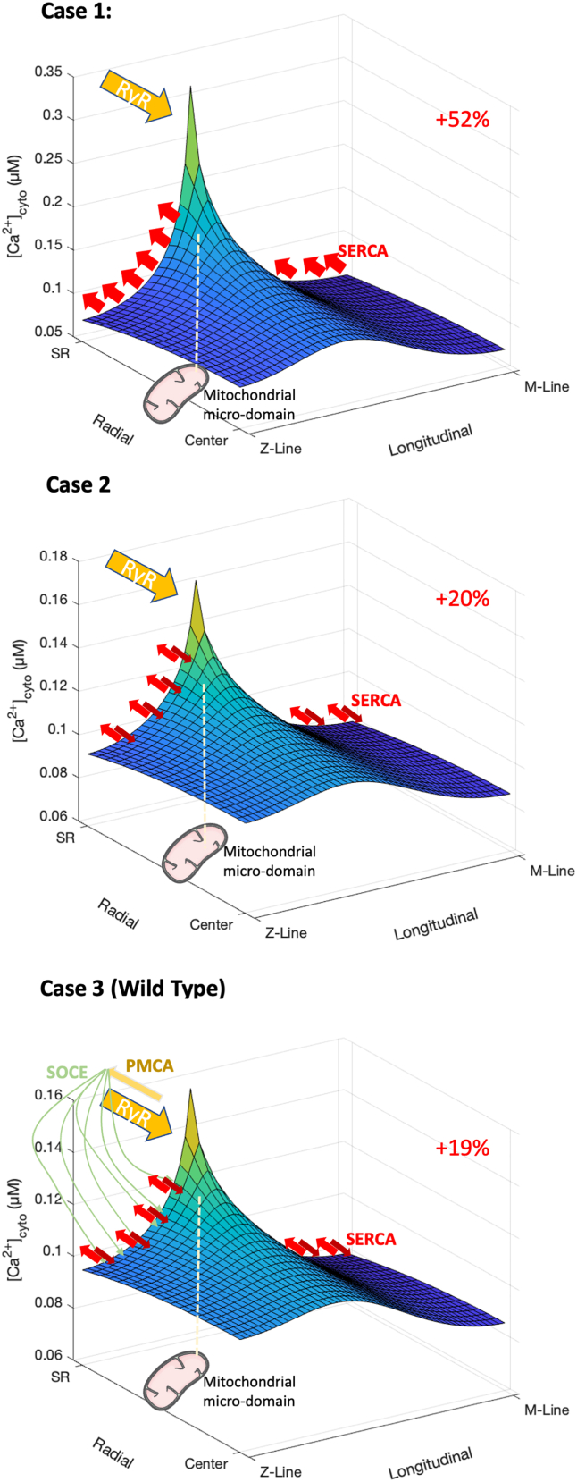
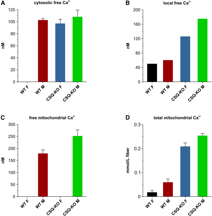
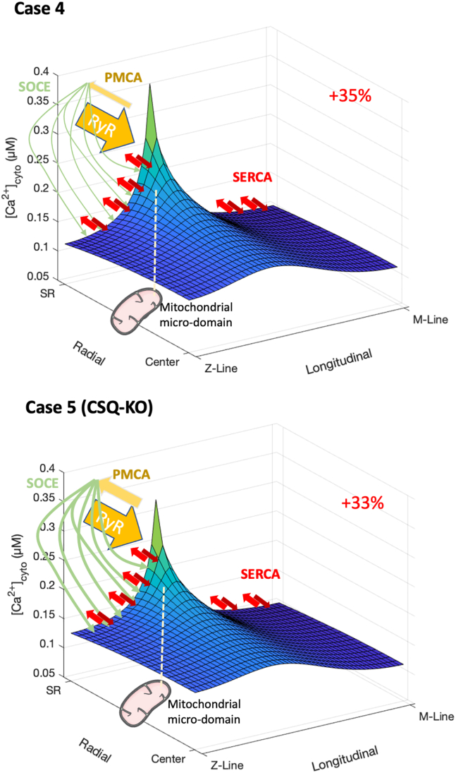
Calcium ions (Ca2+) enter mitochondria via the mitochondrial Ca2+ uniporter, driven by electrical and concentration gradients. In this regard, transgenic mouse models, such as calsequestrin knockout (CSQ-KO) mice, with higher mitochondrial Ca2+ concentrations ([Ca2+]mito), should display higher cytosolic Ca2+ concentrations ([Ca2+]cyto). However, repeated measurements of [Ca2+]cyto in quiescent CSQ-KO fibers never showed a difference between WT and CSQ-KO. Starting from the consideration that fluorescent Ca2+ probes (Fura-2 and Indo-1) measure averaged global cytosolic concentrations, in this report we explored the role of local Ca2+ concentrations (i.e., Ca2+ microdomains) in regulating mitochondrial Ca2+ in resting cells, using a multicompartmental diffusional Ca2+ model. Progressively including the inward and outward fluxes of sarcoplasmic reticulum (SR), extracellular space, and mitochondria, we explored their contribution to the local Ca2+ distribution within the cell. The model predicts Ca2+ concentration gradients with hot spots or microdomains even at rest, minor but similar to those of evoked Ca2+ release. Due to their specific localization close to Ca2+ release units (CRU), mitochondria could take up Ca2+ directly from high-concentration microdomains, thus sensibly raising [Ca2+]mito, despite minor, possibly undetectable, modifications of the average [Ca2+]cyto.


 分享
分享
 求助内容:
求助内容: 应助结果提醒方式:
 扫码关注我们
