Imaging modality-dependent carotid stenosis severity variations against intravascular ultrasound as a reference: Carotid Artery intravasculaR Ultrasound Study (CARUS).
Lukasz Tekieli, Anna Kablak-Ziembicka, Wladyslaw Dabrowski, Karolina Dzierwa, Zbigniew Moczulski, Malgorzata Urbanczyk-Zawadzka, Adam Mazurek, Justyna Stefaniak, Piotr Paluszek, Maciej Krupinski, Tadeusz Przewlocki, Piotr Pieniazek, Piotr Musialek
{"title":"Imaging modality-dependent carotid stenosis severity variations against intravascular ultrasound as a reference: Carotid Artery intravasculaR Ultrasound Study (CARUS).","authors":"Lukasz Tekieli, Anna Kablak-Ziembicka, Wladyslaw Dabrowski, Karolina Dzierwa, Zbigniew Moczulski, Malgorzata Urbanczyk-Zawadzka, Adam Mazurek, Justyna Stefaniak, Piotr Paluszek, Maciej Krupinski, Tadeusz Przewlocki, Piotr Pieniazek, Piotr Musialek","doi":"10.1007/s10554-023-02875-1","DOIUrl":null,"url":null,"abstract":"<p><strong>Purpose: </strong>Different non-invasive and invasive imaging modalities are used to determine carotid artery stenosis severity that remains a principal parameter in clinical decision-making. We compared stenosis degree obtained with different modalities against vascular imaging gold standard, intravascular ultrasound, IVUS.</p><p><strong>Methods: </strong>300 consecutive patients (age 47-83 years, 192 men, 64% asymptomatic) with carotid artery stenosis of \" ≥ 50%\" referred for potential revascularization received as per study protocol (i) duplex ultrasound (DUS), (ii) computed tomography angiography (CTA), (iii) intraarterial quantitative angiography (iQA) and (iv) and (iv) IVUS. Correlation of measurements with IVUS (r), proportion of those concordant (within 10%) and proportion of under/overestimated were calculated along with recipient-operating-characteristics (ROC).</p><p><strong>Results: </strong>For IVUS area stenosis (AS) and IVUS minimal lumen area (MLA), there was only a moderate correlation with DUS velocities (peak-systolic, PSV; end-diastolic, EDV; r values of 0.42-0.51, p < 0.001 for all). CTA systematically underestimated both reference area and MLA (80.4% and 92.3% cases) but CTA error was lesser for AS (proportion concordant-57.4%; CTA under/overestimation-12.5%/30.1%). iQA diameter stenosis (DS) was found concordant with IVUS in 41.1% measurements (iQA under/overestimation 7.9%/51.0%). By univariate model, PSV (ROC area-under-the-curve, AUC, 0.77, cutoff 2.6 m/s), EDV (AUC 0.72, cutoff 0.71 m/s) and CTA-DS (AUC 0.83, cutoff 59.6%) were predictors of ≥ 50% DS by IVUS (p < 0.001 for all). Best predictor, however, of ≥ 50% DS by IVUS was stenosis severity evaluation by automated contrast column density measurement on iQA (AUC 0.87, cutoff 68%, p < 0.001). Regarding non-invasive techniques, CTA was the only independent diagnostic modality against IVUS on multivariate model (p = 0.008).</p><p><strong>Conclusion: </strong>IVUS validation shows significant imaging modality-dependent variations in carotid stenosis severity determination.</p>","PeriodicalId":50332,"journal":{"name":"International Journal of Cardiovascular Imaging","volume":" ","pages":"1909-1920"},"PeriodicalIF":1.5000,"publicationDate":"2023-10-01","publicationTypes":"Journal Article","fieldsOfStudy":null,"isOpenAccess":false,"openAccessPdf":"https://www.ncbi.nlm.nih.gov/pmc/articles/PMC10589130/pdf/","citationCount":"1","resultStr":null,"platform":"Semanticscholar","paperid":null,"PeriodicalName":"International Journal of Cardiovascular Imaging","FirstCategoryId":"3","ListUrlMain":"https://doi.org/10.1007/s10554-023-02875-1","RegionNum":4,"RegionCategory":"医学","ArticlePicture":[],"TitleCN":null,"AbstractTextCN":null,"PMCID":null,"EPubDate":"2023/8/21 0:00:00","PubModel":"Epub","JCR":"Q3","JCRName":"CARDIAC & CARDIOVASCULAR SYSTEMS","Score":null,"Total":0}
引用次数: 1
Abstract
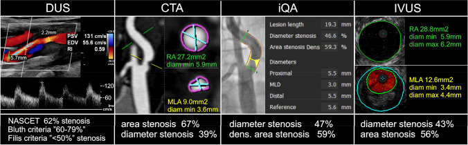
Purpose: Different non-invasive and invasive imaging modalities are used to determine carotid artery stenosis severity that remains a principal parameter in clinical decision-making. We compared stenosis degree obtained with different modalities against vascular imaging gold standard, intravascular ultrasound, IVUS.
Methods: 300 consecutive patients (age 47-83 years, 192 men, 64% asymptomatic) with carotid artery stenosis of " ≥ 50%" referred for potential revascularization received as per study protocol (i) duplex ultrasound (DUS), (ii) computed tomography angiography (CTA), (iii) intraarterial quantitative angiography (iQA) and (iv) and (iv) IVUS. Correlation of measurements with IVUS (r), proportion of those concordant (within 10%) and proportion of under/overestimated were calculated along with recipient-operating-characteristics (ROC).
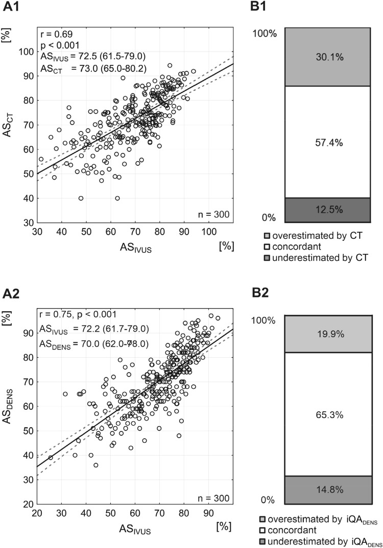
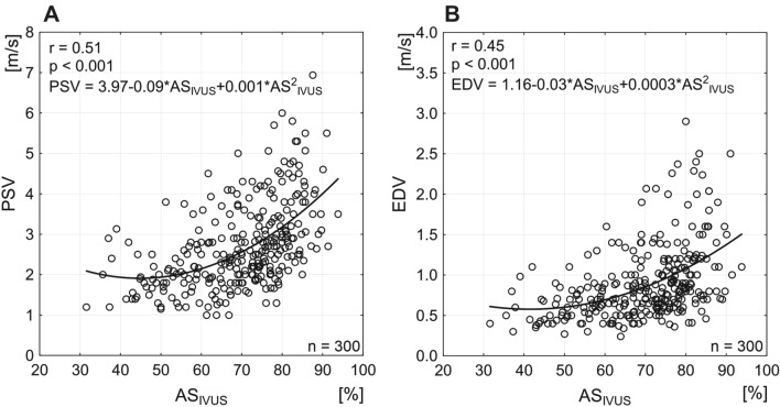
Results: For IVUS area stenosis (AS) and IVUS minimal lumen area (MLA), there was only a moderate correlation with DUS velocities (peak-systolic, PSV; end-diastolic, EDV; r values of 0.42-0.51, p < 0.001 for all). CTA systematically underestimated both reference area and MLA (80.4% and 92.3% cases) but CTA error was lesser for AS (proportion concordant-57.4%; CTA under/overestimation-12.5%/30.1%). iQA diameter stenosis (DS) was found concordant with IVUS in 41.1% measurements (iQA under/overestimation 7.9%/51.0%). By univariate model, PSV (ROC area-under-the-curve, AUC, 0.77, cutoff 2.6 m/s), EDV (AUC 0.72, cutoff 0.71 m/s) and CTA-DS (AUC 0.83, cutoff 59.6%) were predictors of ≥ 50% DS by IVUS (p < 0.001 for all). Best predictor, however, of ≥ 50% DS by IVUS was stenosis severity evaluation by automated contrast column density measurement on iQA (AUC 0.87, cutoff 68%, p < 0.001). Regarding non-invasive techniques, CTA was the only independent diagnostic modality against IVUS on multivariate model (p = 0.008).
期刊介绍:
The International Journal of Cardiovascular Imaging publishes technical and clinical communications (original articles, review articles and editorial comments) associated with cardiovascular diseases. The technical communications include the research, development and evaluation of novel imaging methods in the various imaging domains. These domains include magnetic resonance imaging, computed tomography, X-ray imaging, intravascular imaging, and applications in nuclear cardiology and echocardiography, and any combination of these techniques. Of particular interest are topics in medical image processing and image-guided interventions. Clinical applications of such imaging techniques include improved diagnostic approaches, treatment , prognosis and follow-up of cardiovascular patients. Topics include: multi-center or larger individual studies dealing with risk stratification and imaging utilization, applications for better characterization of cardiovascular diseases, and assessment of the efficacy of new drugs and interventional devices.


 分享
分享
 求助内容:
求助内容: 应助结果提醒方式:
 扫码关注我们
