{"title":"Mucormycosis-related osteomyelitis of the maxilla in a post-COVID-19 patient.","authors":"Yun-Hui Kang, Sam-Sun Lee, Moe Thu Zar Aung, Ju-Hee Kang, Jo-Eun Kim, Kyung-Hoe Huh, Min-Suk Heo","doi":"10.5624/isd.20220143","DOIUrl":null,"url":null,"abstract":"<p><p>Mucormycosis is a rare, invasive fungal infection that progresses aggressively and requires prompt surgery and appropriate treatment. The number of cases of mucormycosis in coronavirus disease 2019 (COVID-19) patients has recently increased, and patients with uncontrolled diabetes mellitus are particularly at an elevated risk of infection. This report presents a case of mucormycosis-related osteomyelitis of the maxilla in a 37-year-old man with diabetes mellitus. The patient complained of severe and persistent pain in the right maxilla, accompanied by increased tooth mobility and headache. On contrast-enhanced computed tomographic images, gas-forming osteomyelitis of the right maxilla was observed. Destruction of the maxilla and palatine bone then proceeded aggressively. Sequestrectomy was performed on the right maxilla, and the histopathological diagnosis was mucormycosis. Further investigation after the first operation revealed the patient's history of COVID-19 infection.</p>","PeriodicalId":51714,"journal":{"name":"Imaging Science in Dentistry","volume":"52 4","pages":"435-440"},"PeriodicalIF":2.1000,"publicationDate":"2022-12-01","publicationTypes":"Journal Article","fieldsOfStudy":null,"isOpenAccess":false,"openAccessPdf":"https://ftp.ncbi.nlm.nih.gov/pub/pmc/oa_pdf/08/9c/isd-52-435.PMC9807792.pdf","citationCount":"0","resultStr":null,"platform":"Semanticscholar","paperid":null,"PeriodicalName":"Imaging Science in Dentistry","FirstCategoryId":"1085","ListUrlMain":"https://doi.org/10.5624/isd.20220143","RegionNum":0,"RegionCategory":null,"ArticlePicture":[],"TitleCN":null,"AbstractTextCN":null,"PMCID":null,"EPubDate":"","PubModel":"","JCR":"Q3","JCRName":"DENTISTRY, ORAL SURGERY & MEDICINE","Score":null,"Total":0}
引用次数: 0
Abstract
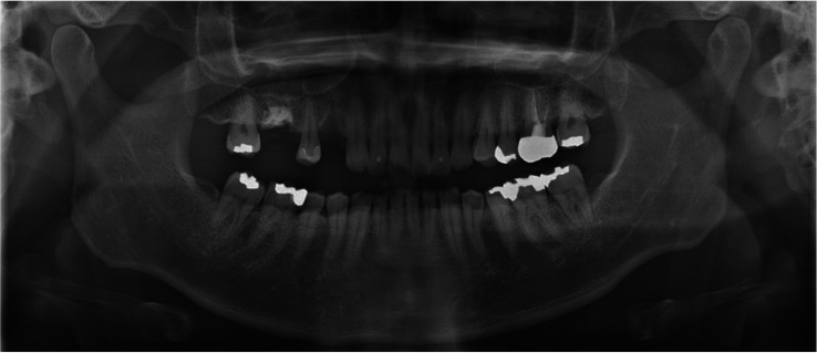
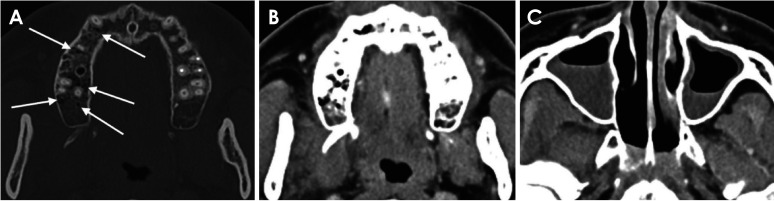
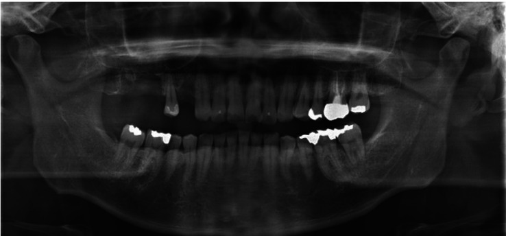
Mucormycosis is a rare, invasive fungal infection that progresses aggressively and requires prompt surgery and appropriate treatment. The number of cases of mucormycosis in coronavirus disease 2019 (COVID-19) patients has recently increased, and patients with uncontrolled diabetes mellitus are particularly at an elevated risk of infection. This report presents a case of mucormycosis-related osteomyelitis of the maxilla in a 37-year-old man with diabetes mellitus. The patient complained of severe and persistent pain in the right maxilla, accompanied by increased tooth mobility and headache. On contrast-enhanced computed tomographic images, gas-forming osteomyelitis of the right maxilla was observed. Destruction of the maxilla and palatine bone then proceeded aggressively. Sequestrectomy was performed on the right maxilla, and the histopathological diagnosis was mucormycosis. Further investigation after the first operation revealed the patient's history of COVID-19 infection.


 分享
分享
 求助内容:
求助内容: 应助结果提醒方式:
 扫码关注我们
